Which Valve Prevents Backflow Into The Left Ventricle
News Leon
Apr 05, 2025 · 6 min read

Table of Contents
Which Valve Prevents Backflow into the Left Ventricle?
The human heart, a marvel of biological engineering, relies on a sophisticated system of valves to ensure unidirectional blood flow. Understanding these valves is crucial for comprehending cardiovascular health and disease. This article delves into the specific valve preventing backflow into the left ventricle, exploring its anatomy, function, and the consequences of its malfunction. We'll also touch upon related conditions and diagnostic procedures.
The Aortic Valve: Guardian of the Left Ventricle
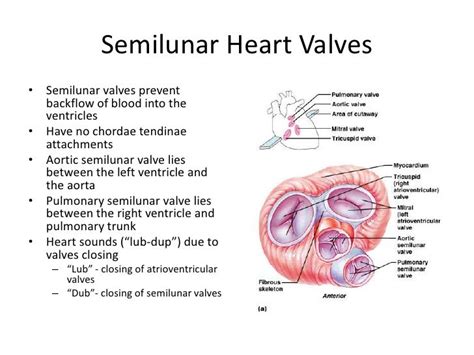
The valve responsible for preventing backflow of blood into the left ventricle is the aortic valve. This crucial valve sits at the exit of the left ventricle, guarding the opening between the ventricle and the aorta, the body's largest artery. Its primary function is to ensure that oxygenated blood, forcefully ejected from the left ventricle during systole (contraction), flows smoothly into the aorta and onwards to the rest of the body. The moment the left ventricle relaxes (diastole), the aortic valve closes tightly, preventing the backflow of blood into the left ventricle.
Anatomy of the Aortic Valve
The aortic valve is a semilunar valve, meaning it's composed of three crescent-shaped leaflets or cusps:
- Right Coronary Cusp: Often the largest cusp, it's positioned closest to the right coronary artery.
- Left Coronary Cusp: Located near the left coronary artery, it's typically the next largest.
- Non-Coronary Cusp: The smallest cusp, it doesn't directly associate with a major coronary artery.
These cusps are thin, but strong and flexible, made of dense connective tissue covered by a thin layer of endothelium (the inner lining of blood vessels). During ventricular contraction, the pressure difference between the left ventricle and the aorta pushes the cusps open, allowing blood to flow freely. As the ventricle relaxes, the pressure in the aorta exceeds that of the ventricle, causing the cusps to close tightly, preventing regurgitation.
Physiology of Aortic Valve Function
The precise timing and efficient closure of the aortic valve are vital for maintaining healthy blood pressure and preventing cardiac strain. The process is tightly regulated by the intricate interplay of pressure gradients and ventricular contraction:
-
Ventricular Systole (Contraction): As the left ventricle contracts, the pressure within rises sharply. This pressure overcomes the pressure in the aorta, forcing the aortic valve cusps open. Blood is rapidly ejected into the aorta.
-
Ventricular Diastole (Relaxation): Once ventricular contraction ceases, the pressure within the left ventricle falls. The higher pressure in the aorta now forces the aortic valve cusps together, creating a tight seal that prevents backflow.
-
Closure and Prevention of Regurgitation: The efficient closure of the aortic valve is critical. Incomplete closure leads to aortic regurgitation, a condition where oxygenated blood flows back into the left ventricle during diastole, reducing the efficiency of the heart.
Consequences of Aortic Valve Dysfunction
Problems with the aortic valve can significantly impact cardiovascular health, leading to various complications:
-
Aortic Stenosis: This condition involves a narrowing of the aortic valve opening, obstructing blood flow from the left ventricle to the aorta. Symptoms may include chest pain (angina), shortness of breath, and dizziness. Severe stenosis can lead to left ventricular hypertrophy (enlargement) and heart failure.
-
Aortic Regurgitation: As mentioned earlier, this refers to the backflow of blood from the aorta into the left ventricle during diastole. It can cause the left ventricle to work harder, leading to enlargement and eventually heart failure. Symptoms often include shortness of breath, chest pain, and a rapid heartbeat.
-
Aortic Valve Disease: This encompasses both stenosis and regurgitation, and may be caused by congenital defects, rheumatic fever, age-related degeneration, or bicuspid aortic valve (a valve with only two cusps instead of three).
Diagnosing Aortic Valve Problems
Several diagnostic tools are available to assess the function of the aortic valve:
-
Echocardiogram (ECHO): This non-invasive ultrasound test produces detailed images of the heart, allowing doctors to visualize the structure and function of the aortic valve, detecting stenosis, regurgitation, or other abnormalities.
-
Cardiac Catheterization: A more invasive procedure involving the insertion of a thin catheter into a blood vessel to reach the heart. This allows for precise measurements of pressure gradients across the aortic valve and assessment of blood flow.
-
Electrocardiogram (ECG or EKG): This test measures the electrical activity of the heart, which can reveal changes associated with aortic valve disease, like left ventricular hypertrophy.
-
Chest X-Ray: While less specific than other tests, a chest X-ray can show signs of left ventricular enlargement, which can indicate aortic valve problems.
Treatment Options for Aortic Valve Disease
Treatment for aortic valve disease depends on the severity of the condition and the patient's overall health:
-
Medication: For mild cases of aortic stenosis or regurgitation, medication may help manage symptoms and slow the progression of the disease. These may include diuretics (to reduce fluid buildup), ACE inhibitors (to control blood pressure), or beta-blockers (to slow the heart rate).
-
Valve Repair or Replacement: For more severe cases, surgical intervention may be necessary. Aortic valve repair aims to correct the problem without completely replacing the valve. However, if repair isn't feasible, aortic valve replacement may be performed, using either a biological valve (from a human or animal donor) or a mechanical valve (made of durable synthetic materials).
-
Transcatheter Aortic Valve Replacement (TAVR): A minimally invasive procedure where a new valve is inserted through a catheter, avoiding the need for open-heart surgery. TAVR is an increasingly popular option for high-risk patients.
Related Cardiovascular Conditions
Understanding the aortic valve's role also requires understanding how its dysfunction can interact with other cardiovascular conditions:
-
Coronary Artery Disease (CAD): Blockages in the coronary arteries that supply blood to the heart muscle. Aortic valve disease can exacerbate CAD, and vice-versa, as the increased workload on the heart due to valve dysfunction can increase the risk of CAD.
-
Heart Failure: The inability of the heart to pump enough blood to meet the body's needs. Aortic valve disease is a common cause of heart failure, as the added strain on the left ventricle eventually leads to its impairment.
-
Hypertension (High Blood Pressure): Chronic high blood pressure can worsen aortic valve disease, especially stenosis, by increasing the strain on the valve. Conversely, severe aortic stenosis can contribute to hypertension.
Conclusion: The Aortic Valve's Crucial Role
The aortic valve plays a crucial role in maintaining the efficient flow of oxygenated blood throughout the body. Its flawless function is essential for overall cardiovascular health. Understanding its anatomy, physiology, and potential for dysfunction is paramount in preventing and treating related cardiovascular conditions. Early diagnosis through appropriate testing and timely intervention, whether through medication or surgery, are key to maintaining a healthy heart and improving quality of life. Regular checkups, especially for individuals with risk factors for aortic valve disease, are highly recommended. By understanding the critical role of the aortic valve, we can better appreciate the remarkable complexity and delicate balance of the human cardiovascular system.
Latest Posts
Latest Posts
-
Which Of The Following Equations Are Identities
Apr 05, 2025
-
Sugar Dissolved In Water Physical Or Chemical Change
Apr 05, 2025
-
12 Is 75 Of What Number
Apr 05, 2025
-
Ammonium Chloride Formula By Criss Cross Method
Apr 05, 2025
-
Is Sr Oh 2 An Acid Or Base
Apr 05, 2025
Related Post
Thank you for visiting our website which covers about Which Valve Prevents Backflow Into The Left Ventricle . We hope the information provided has been useful to you. Feel free to contact us if you have any questions or need further assistance. See you next time and don't miss to bookmark.
