Which Type Of Bacteria Is Shown In The Image
News Leon
Mar 18, 2025 · 5 min read
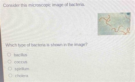
Table of Contents
Identifying Bacteria in Images: A Comprehensive Guide
Determining the precise type of bacteria shown in an image requires a multifaceted approach, combining visual analysis with other crucial factors. While a simple image might not yield a definitive species identification, it can often narrow down the possibilities significantly. This article delves into the methods and considerations involved in bacterial identification from images, focusing on what features to look for and the limitations of relying solely on visual inspection.
The Challenges of Visual Bacterial Identification
Microscopic images of bacteria can be incredibly diverse, depending on factors like:
- Magnification: Low-magnification images might only show colony morphology (shape, size, texture, color), providing little information about individual bacterial cells. Higher magnifications reveal cellular structure, but might not show enough detail for species-level identification.
- Staining techniques: Different staining methods (Gram staining, acid-fast staining, etc.) highlight different bacterial features. A Gram-positive bacterium will appear very differently than a Gram-negative one, even if they're both cocci (spherical). The staining method used profoundly impacts the image's appearance.
- Image quality: Resolution, clarity, and artifacts can significantly impact the ability to identify bacteria. Poorly focused or blurry images render detailed analysis impossible.
- Growth conditions: Bacterial morphology can change based on growth medium, temperature, and other environmental factors. A bacterium grown under one set of conditions might appear different from the same species grown under different conditions.
Key Visual Features for Bacterial Identification
Despite these challenges, several visual characteristics can provide valuable clues when identifying bacteria from images:
1. Cell Shape and Arrangement:
- Cocci (spherical): These can appear singly, in pairs (diplococci), chains (streptococci), clusters (staphylococci), or tetrads (groups of four). The arrangement is a crucial identifying feature.
- Bacilli (rod-shaped): Bacilli can be short and plump, long and slender, or even club-shaped. They can also occur singly, in pairs (diplobacilli), chains (streptobacilli), or palisades (side-by-side arrangements).
- Spirilla (spiral-shaped): These are further categorized into vibrios (comma-shaped), spirilla (rigid spirals), and spirochetes (flexible spirals).
- Coccobacilli: These are bacteria that are intermediate between cocci and bacilli, having a short rod shape that is almost spherical.
2. Gram Staining:
Gram staining is a crucial differential staining technique that divides bacteria into two broad groups based on cell wall composition:
- Gram-positive: Appear purple or blue under a microscope. Their cell walls contain a thick peptidoglycan layer.
- Gram-negative: Appear pink or red. Their cell walls have a thinner peptidoglycan layer and an outer membrane.
3. Spore Formation:
Some bacteria form endospores, highly resistant structures that allow the bacteria to survive harsh conditions. The presence, location (terminal, subterminal, central), and shape of endospores are important identifying characteristics.
4. Capsule:
Some bacteria have a protective layer called a capsule surrounding the cell wall. The presence and appearance of the capsule (size, shape, and staining characteristics) can be diagnostically significant.
5. Flagella:
Flagella are whip-like appendages used for motility. The number and arrangement of flagella (monotrichous, amphitrichous, lophotrichous, peritrichous) are helpful for identification.
6. Pili/Fimbriae:
These are hair-like appendages shorter than flagella, involved in adhesion and conjugation. While harder to visualize than flagella, their presence can be significant.
7. Colony Morphology (Macroscopic Features):
Even before microscopic examination, the colony's macroscopic features offer clues:
- Size: Small, medium, or large colonies.
- Shape: Circular, irregular, filamentous, rhizoid.
- Edge/Margin: Entire (smooth), undulate (wavy), lobate (lobed), erose (scalloped), filamentous.
- Elevation: Flat, raised, convex, umbonate (raised in the center).
- Texture: Smooth, rough, mucoid, wrinkled.
- Pigmentation: Colorless, white, yellow, red, etc.
Limitations of Visual Identification Alone
While the visual characteristics described above provide valuable clues, relying solely on images for bacterial identification is insufficient and potentially inaccurate. Microscopic examination should always be coupled with other tests, including:
- Biochemical tests: These tests determine a bacterium's metabolic capabilities, helping differentiate closely related species. Examples include catalase tests, oxidase tests, and various sugar fermentation tests.
- Molecular techniques: Techniques like 16S rRNA gene sequencing provide highly accurate identification down to the species level. This is often considered the gold standard for bacterial identification.
- Antibiotic susceptibility testing: This helps determine the effectiveness of different antibiotics against the bacterium, providing crucial information for treatment.
- Immunological tests: These tests utilize antibodies to detect specific bacterial antigens.
Example Scenarios and Interpretations:
Let's consider some hypothetical scenarios to illustrate the limitations and possibilities:
Scenario 1: An image shows Gram-positive cocci in chains. This suggests Streptococcus species, but many Streptococcus species exist. Further biochemical tests (e.g., hemolysis patterns on blood agar) are needed to narrow it down.
Scenario 2: The image reveals Gram-negative bacilli with peritrichous flagella. This points towards Escherichia coli or other Enterobacteriaceae, but many species within this family have similar morphologies. Biochemical tests and molecular methods would be required for definite identification.
Scenario 3: A poorly focused image shows a vaguely coccus-shaped organism. Without clarity on Gram staining, arrangement, and other features, any attempt at identification would be highly speculative and unreliable.
Conclusion: A Holistic Approach to Bacterial Identification
Identifying bacteria from images alone is a challenging task. While visual characteristics like cell shape, arrangement, and Gram stain reaction provide important clues, they are insufficient for precise identification. A comprehensive approach combining microscopic observation with biochemical tests, molecular techniques, and possibly immunological methods is necessary to accurately identify bacterial species and ensure reliable results. Remember, the image provides a starting point, not a conclusive answer. Always treat bacterial identification as a multifaceted process requiring multiple lines of evidence.
Latest Posts
Latest Posts
-
A Cell In A Hypertonic Solution Will
Mar 19, 2025
-
What Is 25 Percent Of 25
Mar 19, 2025
-
How Do You Make A Magnet At Home
Mar 19, 2025
-
A Decrease In Demand And An Increase In Supply Will
Mar 19, 2025
-
Prove The Square Root Of 5 Is Irrational
Mar 19, 2025
Related Post
Thank you for visiting our website which covers about Which Type Of Bacteria Is Shown In The Image . We hope the information provided has been useful to you. Feel free to contact us if you have any questions or need further assistance. See you next time and don't miss to bookmark.
