Which Leukocyte Is Responsible For The Allergic Response
News Leon
Mar 25, 2025 · 5 min read
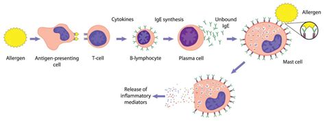
Table of Contents
Which Leukocyte is Responsible for the Allergic Response? The Crucial Role of Mast Cells and Other Immune Players
Allergic reactions, those often uncomfortable and sometimes life-threatening responses to normally harmless substances, are orchestrated by a complex interplay of immune cells. While several leukocytes participate, mast cells are undeniably the central players responsible for the hallmark symptoms of allergic reactions. This article delves deep into the mechanisms of allergic responses, highlighting the crucial role of mast cells, alongside the contributions of other leukocytes like basophils, eosinophils, and lymphocytes.
Understanding Allergic Reactions: A Cascade of Events
An allergic response, also known as a hypersensitivity reaction, is a dysregulated immune reaction to an allergen – a typically harmless antigen like pollen, pet dander, or certain foods. This reaction is characterized by an over-the-top inflammatory response that damages healthy tissues. The process unfolds in a series of phases:
1. Sensitization: The First Encounter
Upon the first exposure to an allergen, the immune system identifies it as foreign. This triggers the activation of B cells, a type of lymphocyte. These B cells differentiate into plasma cells, which then produce IgE antibodies specific to that particular allergen. These IgE antibodies then circulate in the bloodstream.
2. The Mast Cell's Crucial Role: Degranulation and Mediator Release
The next encounter with the same allergen is where the drama begins. This time, the allergen binds to the IgE antibodies that are already pre-bound to the surface receptors of mast cells. This binding triggers degranulation, a process where mast cells release pre-formed mediators stored within their granules. These mediators are the key players in initiating the immediate allergic response.
These mediators include:
-
Histamine: This is the most well-known mediator, causing vasodilation (widening of blood vessels), increased vascular permeability (leakiness of blood vessels), and bronchoconstriction (narrowing of airways). These effects manifest as symptoms such as itching, swelling, redness, and difficulty breathing.
-
Tryptase and Chymase: These proteases further contribute to inflammation and tissue damage.
-
Heparin: This anticoagulant prevents blood clotting.
-
TNF-alpha and IL-4: These cytokines contribute to the amplification of the inflammatory response, recruiting more immune cells to the site of allergen exposure.
The release of these mediators causes the characteristic immediate allergic reactions, which can include:
- Urticaria (hives): Raised, itchy welts on the skin.
- Rhinitis (hay fever): Runny nose, sneezing, itchy eyes.
- Bronchospasm (asthma): Wheezing, coughing, shortness of breath.
- Anaphylaxis: A severe, life-threatening reaction characterized by widespread vasodilation, bronchospasm, and circulatory collapse.
3. Late-Phase Reaction: A Prolonged Inflammatory Response
The immediate reaction is followed by a late-phase reaction, typically occurring hours after the initial exposure. This is a more protracted inflammatory response, driven by the continued release of lipid mediators from mast cells (like leukotrienes and prostaglandins) and the recruitment of other leukocytes, notably eosinophils and basophils. These cells amplify the inflammatory cascade, further contributing to tissue damage and symptoms.
Beyond Mast Cells: The Supporting Cast of Leukocytes
While mast cells are central to the allergic response, other leukocytes contribute significantly to the overall reaction:
Basophils: The Blood-Borne Counterpart to Mast Cells
Basophils are granulocytes found in the bloodstream, sharing many similarities with mast cells, particularly in their expression of IgE receptors and the release of similar mediators (histamine, heparin, leukotrienes). Though less numerous than mast cells at the site of allergic reactions, they contribute to the overall inflammatory response, particularly in the bloodstream. Their role in allergic reactions is thought to be less prominent than that of mast cells, but they amplify the inflammatory cascade.
Eosinophils: Key Players in the Late-Phase Response
Eosinophils are granulocytes primarily involved in the defense against parasites. In allergic reactions, they are recruited to the site of inflammation during the late-phase response. They release a variety of cytotoxic substances, including major basic protein and eosinophil cationic protein, that contribute to tissue damage. While this damage can contribute to the severity of allergic symptoms, eosinophils also play a role in regulating the inflammatory response, potentially limiting its duration. Their increased numbers in allergic diseases like asthma are a key diagnostic marker.
Lymphocytes: Modulators and Amplifiers of the Allergic Cascade
Lymphocytes, particularly T helper cells (Th2 cells), play a crucial role in the development and amplification of allergic inflammation. Th2 cells release cytokines, such as IL-4, IL-5, and IL-13, which promote IgE production by B cells and recruit and activate eosinophils. These cytokines are essential for the chronic inflammation characteristic of many allergic diseases. Regulatory T cells (Tregs) play a counter-regulatory role, trying to suppress the immune response and prevent excessive inflammation. The balance between Th2 and Treg cells is crucial in determining the severity of the allergic response.
Genetic Predisposition and Environmental Factors
The development of allergic diseases is a complex interplay of genetic predisposition and environmental factors. Genetic factors influence the production of IgE antibodies, the responsiveness of mast cells to allergens, and the overall regulation of the immune response. Environmental factors, such as exposure to allergens, pollutants, and infections, can trigger or modulate the allergic response.
Diagnosis and Treatment of Allergic Reactions
Diagnosis of allergic reactions typically involves a detailed history, physical examination, and skin prick tests or blood tests to identify specific allergens. Treatment strategies vary depending on the severity of the reaction and may include avoidance of allergens, antihistamines, corticosteroids, and other medications to control inflammation and symptoms. In severe cases, immunotherapy, also known as allergy shots, may be used to desensitize the immune system to specific allergens. For life-threatening reactions like anaphylaxis, prompt administration of epinephrine is crucial.
Conclusion: A Complex Orchestration of Immune Cells
Allergic responses are not simply the actions of a single cell type but rather a complex, orchestrated response involving many leukocytes. While mast cells are undoubtedly central to the immediate hypersensitivity reaction, initiating the inflammatory cascade and causing many of the hallmark symptoms through the release of potent mediators, basophils, eosinophils, and lymphocytes all contribute significantly to the development and progression of allergic reactions. Understanding the roles of these cells is crucial for the development of effective diagnostic tools and therapeutic strategies to manage allergic diseases. Further research into the intricacies of this complex interplay will undoubtedly lead to improved treatments and potentially preventative measures. This ongoing research focuses on fine-tuning the immune response, targeting specific inflammatory pathways, and developing more effective immunotherapies. The goal remains to effectively manage and ultimately alleviate the burden of allergic diseases on individuals and society.
Latest Posts
Latest Posts
-
Is Boil A Physical Or Chemical Change
Mar 28, 2025
-
Which Mountain Range Separates Europe From Asia
Mar 28, 2025
-
What Was The Poets Childhood Fear
Mar 28, 2025
-
In The Structure Of 4 Isopropyl 2 4 5 Trimethylheptane
Mar 28, 2025
-
Role Of Nad In Cellular Respiration
Mar 28, 2025
Related Post
Thank you for visiting our website which covers about Which Leukocyte Is Responsible For The Allergic Response . We hope the information provided has been useful to you. Feel free to contact us if you have any questions or need further assistance. See you next time and don't miss to bookmark.
