Sister Chromatids Are Joined At The
News Leon
Mar 14, 2025 · 6 min read
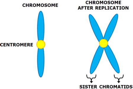
Table of Contents
Sister Chromatids are Joined at the Centromere: A Deep Dive into Chromosome Structure and Function
Sister chromatids are identical copies of a single chromosome that are joined together. This joining occurs at a specific region called the centromere, a crucial structure for accurate chromosome segregation during cell division. Understanding the centromere's structure and function is vital to comprehending the intricacies of the cell cycle and the potential consequences of errors in chromosome segregation. This article will delve into the detailed mechanisms and significance of sister chromatid cohesion at the centromere.
The Structure of the Centromere: A Complex Hub of Activity
The centromere, far from being a simple connection point, is a highly specialized and complex chromosomal region. It's not just a physical linkage; it's a dynamic hub of activity crucial for cell division. Its structure is surprisingly intricate, involving a variety of proteins and DNA sequences that work in concert to ensure proper chromosome segregation.
The Centromeric DNA: More Than Just a Connector
The centromeric DNA itself is not characterized by a specific DNA sequence conserved across all organisms. Instead, it's often characterized by highly repetitive DNA sequences, which can vary significantly even between closely related species. These repetitive sequences are often referred to as satellite DNA due to their distinctive banding patterns in density gradient centrifugation. The abundance of repetitive DNA makes this region challenging to study and understand fully.
However, despite the sequence variability, centromeric DNA plays a crucial role in assembling the kinetochore, the protein structure responsible for attaching the chromosome to the microtubules of the mitotic spindle. Specific DNA sequences within the centromere act as recognition sites for the initial proteins involved in kinetochore formation. These initial proteins then recruit other proteins, eventually leading to the construction of the fully functional kinetochore complex.
The Kinetochore: The Bridge Between Chromosomes and Microtubules
The kinetochore is a complex protein structure assembled on the centromere. It's the crucial interface between the chromosomes and the microtubules of the mitotic spindle. The kinetochore's precise structure and function are essential for accurate chromosome segregation. It has multiple layers, each with specific roles:
-
Inner Kinetochore: This layer directly interacts with the centromeric DNA, binding to specific histone variants and other proteins. This interaction is critical for establishing the initial contact between the chromosome and the mitotic spindle.
-
Outer Kinetochore: This layer interacts with the microtubules of the mitotic spindle. It contains motor proteins and other proteins responsible for the dynamic interactions between the chromosomes and the spindle microtubules. These interactions are responsible for the movement of chromosomes during cell division.
The precise orchestration of protein recruitment and assembly at the kinetochore is essential for the accurate capture and movement of chromosomes during cell division. Errors in this process can lead to aneuploidy—an abnormal number of chromosomes in a cell—which can have severe consequences, including developmental abnormalities and cancer.
Cohesion at the Centromere: Holding Sister Chromatids Together
The joining of sister chromatids at the centromere is mediated by a complex protein complex called cohesin. Cohesin is a ring-shaped protein complex that encircles the two sister chromatids, holding them together. This cohesion is essential for proper chromosome segregation during both mitosis (cell division in somatic cells) and meiosis (cell division in germ cells).
The Role of Cohesin: A Molecular Embrace
Cohesin's role is not simply to physically link the sister chromatids. It actively regulates the timing and location of sister chromatid separation. The loading and removal of cohesin are precisely controlled throughout the cell cycle. Cohesin is loaded onto chromosomes during DNA replication, ensuring that sister chromatids remain tightly associated until the appropriate time for separation.
Cohesin's Regulation: A Tightly Controlled Process
The regulation of cohesin is a complex process involving several factors:
-
Establishment of Cohesion: Cohesin loading occurs during DNA replication, coinciding with the duplication of the chromosomes. Specific proteins facilitate the loading of cohesin onto chromosomes, ensuring that sister chromatids are properly connected.
-
Protection of Cohesion: Once loaded, cohesin must be protected from premature removal. Specific regulatory mechanisms prevent the premature dissociation of cohesin before the appropriate stage of cell division.
-
Regulation of Cohesion Removal: The removal of cohesin is precisely controlled during cell division. A specific enzyme complex, called the anaphase-promoting complex/cyclosome (APC/C), targets cohesin for proteolytic degradation, triggering sister chromatid separation.
The precise regulation of cohesin is essential for ensuring accurate chromosome segregation. Errors in cohesin regulation can lead to premature separation of sister chromatids, resulting in chromosome loss or nondisjunction (failure of chromosomes to separate properly), which can have devastating consequences.
The Significance of Accurate Sister Chromatid Separation
Accurate separation of sister chromatids is paramount for maintaining genome integrity and preventing cellular dysfunction. Errors in this process can have serious consequences:
Aneuploidy: An Imbalance of Chromosomes
Aneuploidy, the presence of an abnormal number of chromosomes in a cell, is a common consequence of errors in sister chromatid separation. This can lead to a range of developmental abnormalities, including birth defects, intellectual disability, and cancer. The severity of aneuploidy depends on which chromosomes are affected and the extent of the imbalance.
Cancer: A Consequence of Genomic Instability
Genomic instability, the increased rate of mutations and chromosomal abnormalities, is a hallmark of cancer. Errors in sister chromatid separation contribute significantly to genomic instability, driving the development and progression of cancer. Chromosomal abnormalities, including aneuploidy, can activate oncogenes (genes that promote cancer development) and inactivate tumor suppressor genes (genes that prevent cancer development).
Developmental Abnormalities: A Spectrum of Effects
Errors in sister chromatid separation during early embryonic development can lead to a wide range of developmental abnormalities. These can vary in severity, depending on the affected chromosomes and the timing of the error. Some abnormalities may be lethal, resulting in miscarriage or stillbirth, while others may cause milder developmental problems.
Research and Future Directions: Unraveling the Mysteries of the Centromere
Research into the structure and function of the centromere and its role in sister chromatid cohesion continues to be an active area of investigation. Several key questions remain unanswered:
-
Centromere Evolution: The mechanisms by which centromeres evolve and the factors that govern the variation in centromeric DNA sequences across species are still not fully understood.
-
Cohesin Regulation: The precise mechanisms regulating cohesin loading, protection, and removal are still under investigation. Further research is needed to fully elucidate the complex regulatory pathways involved.
-
Centromere Dysfunction and Disease: A better understanding of how centromere dysfunction contributes to human diseases, including cancer and developmental abnormalities, is crucial for developing new diagnostic and therapeutic strategies.
Advancements in genomic technologies, including high-throughput sequencing and advanced microscopy techniques, are providing new insights into the intricacies of the centromere and its role in chromosome segregation. These advancements are crucial for furthering our understanding of the fundamental processes of cell division and the consequences of errors in this vital process.
Conclusion: A Vital Process for Life
The joining of sister chromatids at the centromere is a fundamental process for the accurate transmission of genetic information during cell division. The complex interplay of DNA sequences, protein complexes, and regulatory pathways ensures that sister chromatids are held together until the appropriate time for separation. Errors in this process can lead to severe consequences, including aneuploidy, genomic instability, and various diseases. Further research into the intricacies of centromere structure and function is vital for a deeper understanding of cell biology and the development of novel therapies for diseases linked to chromosome segregation errors. The centromere, seemingly a simple connection point, is in fact a remarkably complex and crucial hub of activity, essential for the continued functioning and survival of all eukaryotic life.
Latest Posts
Latest Posts
-
How Many Cubic Centimeters In 1 Cubic Meter
Mar 14, 2025
-
How Many Valence Electrons Does Kr Have
Mar 14, 2025
-
A Flywheel With A Diameter Of 1 20m Is Rotating
Mar 14, 2025
-
Is Al Oh 3 Soluble In Water
Mar 14, 2025
-
The Number Of Protons In An Atom Is Called
Mar 14, 2025
Related Post
Thank you for visiting our website which covers about Sister Chromatids Are Joined At The . We hope the information provided has been useful to you. Feel free to contact us if you have any questions or need further assistance. See you next time and don't miss to bookmark.
