Part Of The Brain That Controls Breathing And Heartbeat
News Leon
Mar 31, 2025 · 6 min read
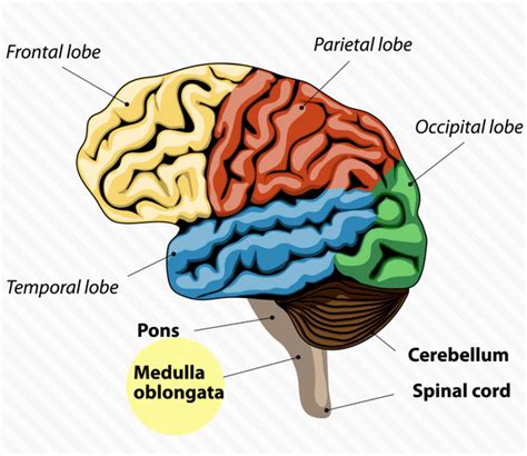
Table of Contents
The Brainstem: The Silent Conductor of Breathing and Heartbeat
The human brain, a marvel of biological engineering, orchestrates the symphony of life. While the cerebrum commands our conscious thoughts and actions, a more primitive yet vital region, the brainstem, silently conducts the fundamental rhythms of existence – breathing and heartbeat. This article delves deep into the intricate neural networks within the brainstem that govern these life-sustaining functions, exploring their anatomy, physiology, and the implications of dysfunction.
The Brainstem: Anatomy and Functional Divisions
The brainstem, the stalk-like structure connecting the cerebrum to the spinal cord, isn't a monolithic entity. Instead, it's composed of three crucial parts: the medulla oblongata, the pons, and the midbrain. Each plays a distinct yet interconnected role in regulating respiratory and cardiovascular functions.
The Medulla Oblongata: The Primary Respiratory and Cardiovascular Control Center
The medulla oblongata, the most caudal (lower) part of the brainstem, houses the vital respiratory and cardiovascular centers. These aren't discrete, easily-identifiable structures but rather diffuse networks of neurons intricately interwoven.
The Respiratory Centers: Rhythm and Response
Within the medulla, two key respiratory centers work in concert:
-
The Dorsal Respiratory Group (DRG): Primarily involved in initiating inspiration (inhaling). It sends signals to the diaphragm and other inspiratory muscles, triggering their contraction. Think of the DRG as the "start button" for each breath.
-
The Ventral Respiratory Group (VRG): Active during both inspiration and expiration (exhaling), the VRG fine-tunes the respiratory rhythm and strength. It contributes to both quiet breathing and forceful breathing during exercise or stress. It can be considered the "modulation center," adjusting the DRG's output based on body demands.
These centers don't operate in isolation. They receive constant input from various sources, including:
-
Chemoreceptors: These specialized cells detect changes in blood oxygen, carbon dioxide, and pH levels. If oxygen levels drop or carbon dioxide rises (leading to acidosis), chemoreceptors signal the respiratory centers to increase breathing rate and depth.
-
Mechanoreceptors: Located in the lungs and airways, these receptors monitor lung stretch and airflow. They prevent overinflation by sending inhibitory signals to the respiratory centers. This is known as the Hering-Breuer reflex.
-
Higher brain centers: Although breathing is largely automatic, conscious control (e.g., holding your breath) can override the brainstem's control. However, the brainstem will ultimately resume control if oxygen levels fall too low.
The Cardiovascular Centers: Heart Rate and Blood Pressure Regulation
The medulla also houses the cardiovascular centers, which precisely regulate heart rate and blood pressure. These include:
-
The Cardioacceleratory Center: Increases heart rate and contractility (the force of heart contractions) through sympathetic nervous system activation. This response is typically triggered by stress, exercise, or low blood pressure.
-
The Cardioinhibitory Center: Slows heart rate through parasympathetic nervous system activation (via the vagus nerve). This center helps maintain a resting heart rate and prevents excessive acceleration.
-
The Vasomotor Center: Controls blood vessel constriction and dilation, thereby influencing blood pressure. It primarily utilizes sympathetic pathways to constrict blood vessels (increasing blood pressure) or dilates them (decreasing blood pressure) as needed.
Like the respiratory centers, the cardiovascular centers receive constant feedback from baroreceptors (pressure sensors in blood vessels) and chemoreceptors, ensuring blood pressure and heart rate remain within the optimal range.
The Pons: Respiratory Rhythm Modulation
The pons, situated above the medulla, doesn't independently control breathing but significantly influences the rhythm and pattern generated by the medullary centers. The pontine respiratory group (PRG) acts as a fine-tuner, smoothing the transitions between inspiration and expiration, preventing abrupt changes in breathing pattern. It's involved in functions like sighing and the deeper breaths we take during sleep.
The Midbrain: Influence on Respiration and Cardiovascular Function
While the midbrain's primary role isn't respiratory or cardiovascular control, it plays a supporting role. It interacts with the lower brainstem centers to integrate respiratory and cardiovascular activity with other bodily functions, particularly during emotional responses such as fear or stress. For example, the midbrain can influence respiratory rate and depth in response to perceived threats.
The Neural Pathways: A Symphony of Signals
The brainstem's control of breathing and heartbeat isn't simply a matter of individual centers acting in isolation. Instead, a complex interplay of neural pathways and neurotransmitters coordinates the finely tuned responses needed to maintain homeostasis.
Sympathetic and Parasympathetic Nervous Systems: The autonomic nervous system (ANS), consisting of the sympathetic and parasympathetic branches, plays a crucial role. The sympathetic nervous system typically increases heart rate and contractility and constricts blood vessels in response to stress or activity, while the parasympathetic system has the opposite effect. These opposing forces are precisely balanced to maintain cardiovascular stability. Similar mechanisms operate for respiratory regulation.
Neurotransmitters: Various neurotransmitters, including acetylcholine, norepinephrine, and serotonin, act as chemical messengers between neurons within the brainstem centers and throughout the autonomic nervous system. Their precise interplay modulates the strength and timing of respiratory and cardiovascular responses.
Feedback Loops: Negative feedback loops are crucial. For example, if blood pressure falls, baroreceptors send signals to the brainstem, triggering a sympathetic response to increase heart rate and blood vessel constriction, bringing pressure back to normal. This constant monitoring and adjustment ensures stability.
Dysfunction and Disorders: When the Silent Conductor Fails
Disruptions to the brainstem's control of breathing and heartbeat can have catastrophic consequences.
Respiratory Disorders:
-
Central Sleep Apnea: Disruptions to the respiratory centers in the medulla cause repeated pauses in breathing during sleep.
-
Ondine's Curse (Congenital Central Hypoventilation Syndrome): A rare genetic disorder characterized by an inability to automatically regulate breathing during sleep. Patients often require mechanical ventilation.
-
Brainstem Stroke: Damage to the medulla or pons can severely impair breathing, potentially leading to respiratory failure.
Cardiovascular Disorders:
-
Cardiac Arrhythmias: Problems with the cardiovascular centers can lead to irregular heartbeats (arrhythmias), including bradycardia (slow heart rate) or tachycardia (fast heart rate).
-
Orthostatic Hypotension: The inability to maintain blood pressure when standing, often due to dysfunction in the autonomic nervous system.
-
Brainstem Stroke: Damage to the medulla can disrupt blood pressure regulation, potentially leading to dangerously high or low blood pressure.
Diagnostic Tools and Treatments
Diagnosing brainstem dysfunction requires a multi-faceted approach. Electroencephalography (EEG), electrocardiography (ECG), polysomnography (sleep study), and blood gas analysis are often employed to assess brain activity, heart rhythm, breathing patterns, and blood oxygen levels.
Treatment varies depending on the underlying condition. For sleep apnea, continuous positive airway pressure (CPAP) therapy may be used. Cardiac arrhythmias may require medication or implantable devices (pacemakers or defibrillators). Respiratory failure may necessitate mechanical ventilation. In some cases, surgery may be an option to address structural abnormalities or repair damaged tissues.
Conclusion: The Unsung Hero
The brainstem, often overlooked in discussions of brain function, is an unsung hero. Its silent control of breathing and heartbeat is the foundation upon which all other functions depend. Understanding the intricate anatomy, physiology, and potential points of failure within this vital region is crucial for advancing medical diagnostics and therapies related to life-threatening respiratory and cardiovascular conditions. Further research into the complex interactions within the brainstem's neural networks promises to provide deeper insights into maintaining the delicate balance that sustains life.
Latest Posts
Latest Posts
-
Balanced Equation For Combustion Of Ethane
Apr 01, 2025
-
Every Integer Is A Real Number
Apr 01, 2025
-
Count Vowels In A String Python
Apr 01, 2025
-
Which Of The Following Elements Is Most Electronegative
Apr 01, 2025
-
For Which Value Of X Is Abcd A Kite
Apr 01, 2025
Related Post
Thank you for visiting our website which covers about Part Of The Brain That Controls Breathing And Heartbeat . We hope the information provided has been useful to you. Feel free to contact us if you have any questions or need further assistance. See you next time and don't miss to bookmark.
