Draw And Label One Complete Cell Cycle
News Leon
Apr 02, 2025 · 7 min read

Table of Contents
Draw and Label One Complete Cell Cycle: A Comprehensive Guide
The cell cycle is a fundamental process in all living organisms, representing the series of events that lead to cell growth and division. Understanding the cell cycle is crucial for grasping the intricacies of life itself, from development to disease. This comprehensive guide will delve into the intricacies of the cell cycle, providing a detailed description, visual representation, and explanations of each stage. We will also explore the regulatory mechanisms that ensure the fidelity of this crucial process.
The Phases of the Cell Cycle: A Detailed Breakdown
The cell cycle is broadly divided into two major phases: interphase and the M phase (mitotic phase). Interphase, the longest phase, prepares the cell for division, while the M phase encompasses the actual division process. Let's examine each phase in detail:
Interphase: The Preparatory Phase
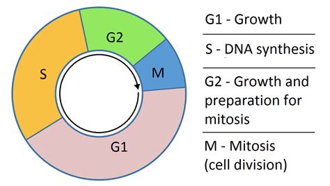
Interphase is further subdivided into three stages:
1. G1 (Gap 1) Phase: This is a period of intense cellular growth and metabolic activity. The cell increases in size, synthesizes proteins and organelles, and performs its specialized functions. This is also a critical checkpoint where the cell assesses its readiness to proceed to DNA replication. If conditions are unfavorable (e.g., nutrient deprivation, DNA damage), the cell may enter a non-dividing state called G0.
2. S (Synthesis) Phase: During this stage, DNA replication occurs. Each chromosome is duplicated, creating two identical sister chromatids joined at the centromere. This ensures that each daughter cell receives a complete set of genetic information. The centrosome, the microtubule-organizing center, is also duplicated during this phase.
3. G2 (Gap 2) Phase: This phase is characterized by further cell growth and preparation for mitosis. The cell checks for any errors in DNA replication and makes necessary repairs. The cell also synthesizes proteins needed for mitosis, such as microtubules. Another checkpoint ensures the cell is ready to proceed to mitosis.
M Phase: The Division Phase
The M phase consists of two main processes: mitosis and cytokinesis.
1. Mitosis: Mitosis is the process of nuclear division, resulting in the equal distribution of replicated chromosomes into two daughter nuclei. It is further divided into several distinct stages:
-
Prophase: Chromosomes condense and become visible under a microscope. The nuclear envelope begins to break down, and the mitotic spindle, a structure made of microtubules, starts to form between the centrosomes, which have migrated to opposite poles of the cell.
-
Prometaphase: The nuclear envelope completely fragments. Kinetochores, protein structures at the centromeres of chromosomes, attach to the microtubules of the spindle. Chromosomes begin to move toward the metaphase plate.
-
Metaphase: Chromosomes align at the metaphase plate, an imaginary plane equidistant from the two poles of the spindle. This alignment ensures that each daughter cell will receive one copy of each chromosome. This stage is crucial for accurate chromosome segregation.
-
Anaphase: Sister chromatids separate at the centromeres and move toward opposite poles of the spindle. This separation is driven by the shortening of the microtubules.
-
Telophase: Chromosomes arrive at the poles and begin to decondense. The nuclear envelope reforms around each set of chromosomes, creating two separate nuclei. The mitotic spindle disassembles.
2. Cytokinesis: This is the final stage of the cell cycle, where the cytoplasm divides, resulting in two separate daughter cells. In animal cells, a cleavage furrow forms, pinching the cell in two. In plant cells, a cell plate forms between the two nuclei, eventually developing into a new cell wall.
Visual Representation: Drawing and Labeling the Cell Cycle
To truly understand the cell cycle, visualizing it is crucial. Below is a description of how to draw and label a complete cell cycle diagram:
1. Interphase:
- Draw a large circle to represent the cell.
- Label the nucleus within the circle.
- Divide the circle into three smaller sections, representing G1, S, and G2.
- Within the G1 section, draw a few small circles and lines to represent organelles and cellular components. Label this as G1 (Gap 1): Cell Growth.
- In the S section, draw the same structures as in G1 but with each chromosome duplicated, represented by an "X" shape. Label this as S (Synthesis): DNA Replication.
- In the G2 section, show slightly larger structures, indicating further growth. Label this as G2 (Gap 2): Preparation for Mitosis. You might include duplicated centrosomes.
2. M Phase (Mitosis):
- Draw a series of circles to represent the stages of mitosis, connected to the G2 section.
- Prophase: Show the chromosomes condensing and thickening, and the nuclear envelope starting to break down. Label the chromosomes and the forming spindle. Label this section as Prophase: Chromosome Condensation.
- Prometaphase: Show the nuclear envelope completely broken down, chromosomes attaching to the spindle fibers at their kinetochores. Label this section as Prometaphase: Chromosome Attachment.
- Metaphase: Show the chromosomes aligned at the metaphase plate. Label the metaphase plate, chromosomes, and spindle fibers. Label this section as Metaphase: Chromosome Alignment.
- Anaphase: Show the sister chromatids separating and moving toward opposite poles. Label the separating chromatids and the spindle fibers. Label this section as Anaphase: Sister Chromatid Separation.
- Telophase: Show the chromosomes at the poles, decondensed, and the nuclear envelopes reforming. Label the new nuclei and chromosomes. Label this section as Telophase: Nuclear Envelope Reformation.
- Cytokinesis: Draw two smaller circles, separated from the telophase circles. Label this section as Cytokinesis: Cytoplasmic Division. Show a cleavage furrow (animal cell) or cell plate (plant cell) forming.
3. Labeling:
Clearly label all parts of your diagram, including:
- G1, S, G2: The phases of interphase.
- Prophase, Prometaphase, Metaphase, Anaphase, Telophase: The phases of mitosis.
- Cytokinesis: The final division of the cytoplasm.
- Chromosomes: Clearly indicate individual chromosomes and sister chromatids.
- Centromere: The point where sister chromatids are joined.
- Spindle Fibers: The microtubules that pull the chromosomes apart.
- Nuclear Envelope: The membrane surrounding the nucleus.
- Centrosomes: The microtubule-organizing centers.
- Cleavage Furrow/Cell Plate: The structures involved in cytokinesis.
Regulatory Mechanisms: Ensuring Accurate Cell Division
The cell cycle is tightly regulated by a complex network of proteins called cyclins and cyclin-dependent kinases (CDKs). These molecules act as checkpoints, ensuring that each stage of the cycle is completed accurately before proceeding to the next. These checkpoints monitor for DNA damage, proper chromosome replication, and accurate spindle attachment. If errors are detected, the cycle is halted, allowing for repair or programmed cell death (apoptosis). Several key checkpoints exist:
- G1 Checkpoint: This is a critical checkpoint that determines whether the cell proceeds to DNA replication. It monitors cell size, nutrient availability, and DNA damage.
- G2 Checkpoint: This checkpoint ensures that DNA replication is complete and accurate before the cell enters mitosis.
- Metaphase Checkpoint: This checkpoint, also known as the spindle checkpoint, ensures that all chromosomes are properly attached to the spindle before anaphase begins.
Errors in the Cell Cycle and Their Consequences
Errors in the cell cycle can have dire consequences, leading to various diseases, including cancer. Uncontrolled cell division, a hallmark of cancer, is often caused by mutations in genes that regulate the cell cycle. These mutations can lead to the bypassing of checkpoints, resulting in the proliferation of cells with damaged DNA.
Conclusion
The cell cycle is a remarkable process, essential for the growth and reproduction of all living organisms. Understanding its intricate details, including the phases, regulatory mechanisms, and potential consequences of errors, is vital for advancing our knowledge in fields such as medicine, genetics, and developmental biology. By accurately drawing and labeling a complete cell cycle, we can effectively visualize and comprehend this fundamental biological process. The comprehensive representation detailed above allows for a thorough understanding of each phase and the critical checkpoints that maintain the fidelity of cell division. This visual aid serves as a valuable tool for learning and understanding this complex yet essential process. Remember to always practice and refine your diagrams to improve your understanding and mastery of this crucial biological concept.
Latest Posts
Latest Posts
-
Do Annelids Have An Open Or Closed Circulatory System
Apr 03, 2025
-
Which Is Not A Component Of Emotional Intelligence
Apr 03, 2025
-
Arrange The Following Radicals In Order Of Increasing Stability
Apr 03, 2025
-
The Programmer Usually Enters Source Code Into A Computer With
Apr 03, 2025
-
When Dissolved In Water Acids Produce
Apr 03, 2025
Related Post
Thank you for visiting our website which covers about Draw And Label One Complete Cell Cycle . We hope the information provided has been useful to you. Feel free to contact us if you have any questions or need further assistance. See you next time and don't miss to bookmark.
