Dendrites Differ From Axons In That Dendrites
News Leon
Mar 20, 2025 · 7 min read
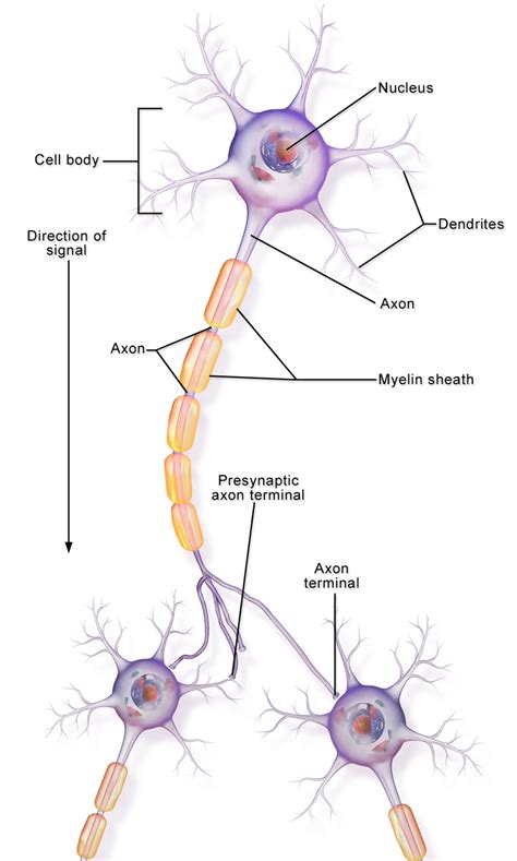
Table of Contents
Dendrites vs. Axons: Key Differences in Neuronal Structure and Function
Neurons, the fundamental units of the nervous system, are responsible for receiving, processing, and transmitting information throughout the body. This complex communication relies heavily on the intricate structure of the neuron itself, particularly the distinct roles played by dendrites and axons. While both are crucial for neuronal function, dendrites and axons exhibit several key differences in their structure, function, and neurochemical properties. Understanding these differences is essential for comprehending how the nervous system works and how neurological disorders can arise from disruptions in neuronal communication.
The Morphology of Dendrites and Axons: A Structural Comparison
The most immediate distinction between dendrites and axons lies in their morphology. Dendrites, typically numerous and branching, resemble a tree-like structure extending from the neuron's soma (cell body). Their branching pattern, known as dendritic arborization, is highly variable depending on the neuron type and location in the nervous system. This extensive branching vastly increases the surface area available for receiving synaptic inputs from other neurons. In contrast, axons usually arise as a single, relatively long projection from the soma, known as the axon hillock. While axons can also branch, forming axon collaterals, their branching is typically less extensive than that of dendrites.
Dendritic Spines: Sites of Synaptic Input
A further structural difference lies in the presence of dendritic spines. These small, protrusions along the dendrites are the primary sites of synaptic input. The morphology of dendritic spines is highly dynamic, changing in size and shape in response to synaptic activity. This plasticity is thought to play a critical role in learning and memory. Axons, on the other hand, lack dendritic spines; they transmit signals away from the soma, not receiving them directly as dendrites do.
Myelin Sheath and Nodes of Ranvier: A Feature of Axons
Axons, particularly those involved in long-distance signaling, are often covered by a myelin sheath. This insulating layer, formed by glial cells (oligodendrocytes in the central nervous system and Schwann cells in the peripheral nervous system), dramatically increases the speed of action potential propagation. The myelin sheath is interrupted at regular intervals by Nodes of Ranvier, which are unmyelinated gaps where ion channels are concentrated. This arrangement facilitates saltatory conduction, a process where action potentials "jump" between nodes, leading to faster and more efficient signal transmission. Dendrites, conversely, generally lack a myelin sheath and conduct signals passively, meaning that the signal strength degrades with distance from the synapse.
Functional Differences: Signal Transmission and Integration
The structural differences between dendrites and axons are directly related to their distinct functional roles. Dendrites are primarily responsible for receiving signals from other neurons. Neurotransmitters released from presynaptic terminals bind to receptors on the dendritic membrane, triggering changes in the membrane potential. These postsynaptic potentials can be either excitatory (EPSPs), depolarizing the membrane and making it more likely to fire an action potential, or inhibitory (IPSPs), hyperpolarizing the membrane and making it less likely to fire. Dendrites then integrate these numerous synaptic inputs, summing them both spatially (across multiple synapses) and temporally (over time).
Axons, on the other hand, are responsible for transmitting signals away from the soma to other neurons, muscles, or glands. When the summated postsynaptic potentials at the axon hillock reach a threshold, an action potential is generated. This action potential is a rapid, all-or-nothing electrical signal that propagates along the axon to its terminals.
Neurotransmitter Release at Axon Terminals
At the axon terminals, or synaptic boutons, the arrival of the action potential triggers the release of neurotransmitters into the synaptic cleft. These neurotransmitters then diffuse across the cleft and bind to receptors on the postsynaptic neuron's dendrites, initiating a new cycle of signal transmission. Dendrites do not release neurotransmitters in this manner; their role is purely receptive.
Molecular Differences: Protein Expression and Membrane Composition
The contrasting functions of dendrites and axons are reflected in the distinct molecular composition of their membranes. Different types of ion channels, neurotransmitter receptors, and other membrane proteins are expressed at higher levels in dendrites compared to axons, or vice-versa. For instance, voltage-gated sodium channels, crucial for action potential generation and propagation, are highly concentrated in the axon but are sparsely distributed in dendrites. In contrast, various types of ligand-gated ion channels and G-protein coupled receptors are more abundant in the dendritic membrane, reflecting their role in receiving and integrating synaptic inputs.
The Role of Microtubules and Neurofilaments
The cytoskeleton also plays a critical role in the structural and functional differentiation of dendrites and axons. Microtubules are essential for maintaining the shape and transporting molecules within both dendrites and axons. However, the organization and dynamics of microtubules differ between the two. Axons typically exhibit a more uniform and polarized arrangement of microtubules, crucial for long-distance transport of materials along the axon. Dendrites have a more complex microtubule organization reflecting their more intricate branching and specialized signaling functions. Neurofilaments, another component of the neuronal cytoskeleton, are also more abundant in axons than in dendrites, contributing to the mechanical strength of the axon and its resistance to compression.
Dendritic Integration and Synaptic Plasticity
The integration of synaptic inputs within dendrites is a highly complex process. The dendritic tree is not a simple passive conductor of signals; rather, it actively processes information through various mechanisms, including spatial summation, temporal summation, and dendritic filtering. Spatial summation involves the summing of postsynaptic potentials from multiple synapses at different locations on the dendrite. Temporal summation involves the summing of postsynaptic potentials arriving at the same synapse over time. Dendritic filtering refers to the selective amplification or attenuation of specific signal frequencies based on the dendritic morphology and the distribution of ion channels.
Dendritic integration plays a pivotal role in synaptic plasticity, the ability of synapses to strengthen or weaken over time in response to activity. This plasticity is crucial for learning and memory and involves changes in synaptic strength, receptor expression, and dendritic spine morphology. Long-term potentiation (LTP) and long-term depression (LTD) are two prominent examples of synaptic plasticity mechanisms that are thought to underpin the formation and consolidation of memories.
Axonal Transport and Signal Propagation
Axons rely on sophisticated transport mechanisms to move essential molecules, such as neurotransmitters, proteins, and organelles, between the soma and the axon terminals. This process, known as axonal transport, occurs along microtubules via molecular motors such as kinesin and dynein. Anterograde transport moves materials from the soma to the axon terminals, while retrograde transport moves materials in the opposite direction. Disruptions in axonal transport can have severe consequences, leading to neuronal dysfunction and neurodegenerative diseases.
The propagation of action potentials along the axon is a fundamental process for neuronal communication. In myelinated axons, saltatory conduction allows for rapid and efficient signal transmission. In unmyelinated axons, action potentials propagate continuously along the axon membrane, a slower process. The speed of action potential propagation is determined by several factors, including axon diameter, myelin sheath thickness, and the density of ion channels.
Clinical Significance: Neurological Disorders and Neuronal Dysfunction
Disruptions in the structure and function of dendrites and axons are implicated in a wide range of neurological disorders. For example, impaired dendritic arborization and spine density have been observed in several neurodevelopmental and neurodegenerative diseases, including Alzheimer's disease and schizophrenia. Similarly, axonal damage and demyelination are hallmarks of multiple sclerosis and other neurological conditions. Understanding the specific mechanisms by which these disorders affect dendrites and axons is crucial for developing effective treatments.
Conclusion: A Coordinated Dance of Neuronal Communication
In conclusion, dendrites and axons, although both integral to neuronal function, are fundamentally different in their structure, function, and molecular composition. Dendrites are specialized for receiving and integrating synaptic inputs, while axons are responsible for transmitting signals over long distances. The precise interplay between these two components ensures the efficient and coordinated processing and transmission of information within the nervous system. Disruptions to either component can have devastating consequences, highlighting the importance of maintaining the integrity of both dendrites and axons for proper brain function and preventing neurological disorders. Further research into the intricacies of dendrite-axon interaction promises to shed even more light on the complexities of the nervous system and potentially provide novel therapeutic strategies for neurological diseases.
Latest Posts
Latest Posts
-
What Is The Greatest Common Factor Of 36 And 54
Mar 21, 2025
-
Which Layer Of The Skin Does Not Contain Blood Vessels
Mar 21, 2025
-
How Many Neutrons Does Xenon Have
Mar 21, 2025
-
How Many Light Years Is Pluto From Earth
Mar 21, 2025
-
Sequence Is True For The Lytic Cycle Of A Virus
Mar 21, 2025
Related Post
Thank you for visiting our website which covers about Dendrites Differ From Axons In That Dendrites . We hope the information provided has been useful to you. Feel free to contact us if you have any questions or need further assistance. See you next time and don't miss to bookmark.
