Correctly Label The Following Parts Of A Renal Corpuscle.
News Leon
Mar 15, 2025 · 6 min read
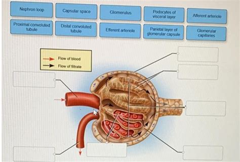
Table of Contents
Correctly Label the Following Parts of a Renal Corpuscle: A Deep Dive into Nephron Structure and Function
The renal corpuscle, the initial filtering unit of the nephron, is a critical structure within the kidney responsible for the intricate process of blood filtration. Understanding its components is fundamental to grasping the mechanics of urine formation and overall renal physiology. This comprehensive guide will delve deep into the renal corpuscle, detailing each part and its function, along with related clinical considerations.
The Renal Corpuscle: A Microscopic Marvel
The renal corpuscle, also known as the Malpighian corpuscle, is the beginning of the nephron, the functional unit of the kidney. It's composed of two main structures: the glomerulus and Bowman's capsule. These structures work in concert to filter blood and initiate the process of urine formation. The precise labeling of these components and their substructures is essential for accurate understanding of renal function.
1. The Glomerulus: A Network of Capillaries
The glomerulus is a tuft of specialized capillaries nestled within Bowman's capsule. It's not just any capillary bed; its structure is uniquely adapted for filtration. Let's break down its key features:
1.1. Glomerular Endothelial Cells: The First Line of Defense
The glomerular capillaries are lined with fenestrated endothelial cells. These cells possess numerous pores or fenestrae, creating a highly permeable barrier. These pores are larger than those found in typical capillaries, allowing for the passage of water and small solutes while largely excluding larger molecules like proteins. However, these fenestrae are not completely open; a glycocalyx layer covers the endothelial surface, adding another layer of filtration selectivity.
1.2. Glomerular Basement Membrane (GBM): A Selective Filter
Surrounding the glomerular endothelial cells is the glomerular basement membrane (GBM). This is a crucial component of the filtration barrier. The GBM is a complex structure composed of three layers: the lamina rara interna (closest to the endothelium), the lamina densa (the middle layer, rich in type IV collagen), and the lamina rara externa (closest to the podocytes). The GBM acts as a size and charge selective filter, restricting the passage of larger molecules and negatively charged proteins. Its intricate structure allows for precise regulation of what passes into Bowman's space.
1.3. Podocytes: The Gatekeepers of Filtration
The outermost layer of the glomerular filtration barrier is formed by podocytes. These specialized epithelial cells have elaborate foot-like processes called pedicels that interdigitate, leaving narrow filtration slits between them. These slits are covered by a thin membrane called the slit diaphragm, containing specialized proteins like nephrin and podocin, which further regulate the passage of molecules. Podocytes play a critical role in regulating glomerular permeability and preventing the passage of large proteins into the filtrate. Damage to podocytes is frequently implicated in proteinuria (protein in the urine).
2. Bowman's Capsule: Enclosing the Glomerulus
Bowman's capsule, also known as the glomerular capsule, is a double-walled epithelial cup that surrounds the glomerulus. It's divided into two layers:
2.1. Parietal Layer: The Outer Layer
The parietal layer of Bowman's capsule is the outer layer, composed of simple squamous epithelium. It forms the structural boundary of the renal corpuscle but doesn't participate directly in filtration. Its primary function is to provide structural support and enclose the filtration apparatus.
2.2. Visceral Layer: The Inner Layer
The visceral layer is the inner layer of Bowman's capsule and directly interacts with the glomerulus. This layer is made up of the specialized podocytes mentioned earlier, whose intricate processes and slit diaphragms play a vital role in the filtration process. The visceral layer is intimately involved in the selective filtration of blood.
2.3. Bowman's Space (Capsular Space): Collecting the Filtrate
Between the parietal and visceral layers lies the Bowman's space, also known as the capsular space. This is the space where the filtrate, the fluid filtered from the blood, collects before entering the proximal convoluted tubule. The composition of the filtrate is significantly different from that of plasma due to the selective filtration by the glomerular filtration barrier.
The Filtration Process: A Symphony of Structure and Function
The glomerular filtration process is driven by hydrostatic pressure differences across the glomerular filtration barrier. The high blood pressure within the glomerular capillaries forces water and small solutes through the porous endothelium, the GBM, and the slit diaphragms of the podocytes into Bowman's space. This filtrate, while containing many of the components of plasma, is largely devoid of large proteins and blood cells. The efficiency of this process is determined by the interplay between hydrostatic and oncotic pressures, and any disruption in the structure of the filtration barrier can significantly affect filtration rates.
Clinical Significance: Understanding Renal Dysfunction
Understanding the anatomy and physiology of the renal corpuscle is paramount for diagnosing and managing various renal diseases. Damage to any component of the filtration barrier can lead to significant alterations in filtration rates and urine composition. For example:
-
Glomerulonephritis: Inflammation of the glomeruli can damage the glomerular capillaries, GBM, and podocytes, leading to proteinuria, hematuria (blood in the urine), and decreased glomerular filtration rate (GFR).
-
Diabetic Nephropathy: High blood glucose levels can damage the glomerular capillaries and GBM, leading to progressive kidney damage and eventual kidney failure.
-
Focal Segmental Glomerulosclerosis (FSGS): This condition involves scarring of parts of the glomeruli, leading to proteinuria and decreased GFR. It's often associated with podocyte injury.
-
Nephrotic Syndrome: A collection of symptoms, including severe proteinuria, hypoalbuminemia (low blood albumin), edema (swelling), and hyperlipidemia (high blood lipids), often results from damage to the glomerular filtration barrier.
Accurate labeling and understanding of the renal corpuscle structures are vital for clinicians to interpret laboratory findings, such as urine analysis and GFR measurements, and to formulate appropriate treatment plans for patients with renal disorders.
Beyond the Renal Corpuscle: The Nephron's Journey
The filtrate collected in Bowman's space continues its journey through the nephron, undergoing further modifications as it flows through the proximal convoluted tubule, loop of Henle, distal convoluted tubule, and collecting duct. These segments are responsible for the reabsorption of essential nutrients, water, and ions, as well as the secretion of waste products into the urine. The precise regulation of these processes is essential for maintaining electrolyte balance, blood pressure, and overall homeostasis.
Conclusion: A Foundation for Renal Understanding
The renal corpuscle, with its intricate architecture and precisely orchestrated filtration mechanisms, forms the cornerstone of renal physiology. Correctly labeling its components—the glomerulus (including its endothelial cells, GBM, and podocytes), Bowman's capsule (parietal and visceral layers), and Bowman's space—provides a fundamental understanding of the process of urine formation and the maintenance of homeostasis. Appreciating its structure and function is crucial for diagnosing and managing a wide range of renal diseases, highlighting the critical role of this microscopic marvel in overall human health. Further investigation into each individual component and its related pathologies opens up a world of understanding in the field of nephrology. Continued research and advancements in this area are vital for improved diagnosis and treatment of kidney diseases globally.
Latest Posts
Latest Posts
-
Which Heart Chamber Has The Thickest Wall
Mar 15, 2025
-
Viral Capsids Are Made From Subunits Called
Mar 15, 2025
-
A Negatively Charged Ion Is Called
Mar 15, 2025
-
Two Different Isotopes Of An Element Have Different
Mar 15, 2025
-
If A Pea Plant Shows A Recessive Phenotype
Mar 15, 2025
Related Post
Thank you for visiting our website which covers about Correctly Label The Following Parts Of A Renal Corpuscle. . We hope the information provided has been useful to you. Feel free to contact us if you have any questions or need further assistance. See you next time and don't miss to bookmark.
