Carries Blood Away From The Kidney
News Leon
Mar 25, 2025 · 7 min read
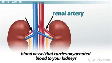
Table of Contents
The Renal Vein: Carrying Blood Away from the Kidney
The kidneys, vital organs responsible for filtering waste products from the blood, rely on a sophisticated vascular system to perform their functions effectively. Understanding this system is key to grasping the intricacies of renal physiology. This article delves into the crucial role of the renal vein, the vessel responsible for carrying filtered blood away from the kidneys back to the heart. We’ll explore its anatomy, function, clinical significance, and the potential consequences of disruptions to its normal operation.
Anatomy of the Renal Vein
The renal vein is a large vein that emerges from the hilum of the kidney—the indented region where the renal artery, renal vein, ureter, and nerves enter and exit the organ. Each kidney possesses its own renal vein, which differs in size and location depending on the individual and the specific kidney. The left and right renal veins, however, exhibit key anatomical distinctions:
Right Renal Vein: A Relatively Straightforward Path
The right renal vein is generally shorter and straighter than its counterpart. It travels directly from the hilum of the right kidney to the inferior vena cava (IVC), the large vein that carries deoxygenated blood from the lower body back to the heart. Its relatively uncomplicated path minimizes potential for obstruction.
Left Renal Vein: A More Complex Journey
The left renal vein has a more complex anatomical course. It is significantly longer than the right renal vein and passes anterior (in front of) the abdominal aorta before joining the IVC. This longer path necessitates its passage behind the superior mesenteric artery (SMA) and often requires it to receive the left gonadal vein (testicular or ovarian vein) and left suprarenal (adrenal) vein before it joins the IVC. This confluence of veins can make the left renal vein more susceptible to compression or obstruction.
Physiology: The Flow of Filtered Blood
The primary function of the renal vein is to transport deoxygenated blood that has been filtered by the kidneys. This filtered blood is now devoid of many waste products and excess fluids, having undergone the processes of glomerular filtration, tubular reabsorption, and tubular secretion within the nephrons, the functional units of the kidney.
The renal vein plays a crucial role in maintaining homeostasis, the body’s internal balance. By efficiently removing waste products and regulating fluid balance, it contributes to the overall health and well-being of the organism. The precise balance of electrolytes, such as sodium, potassium, and chloride, is also influenced by the efficient functioning of the renal vein. Impaired function here can lead to imbalances with serious consequences.
The efficient flow of blood through the renal vein is essential. Any impediment to this flow can lead to a build-up of pressure within the renal veins and the kidneys themselves. This increased pressure can damage the delicate structures within the kidney, leading to a variety of health problems.
Clinical Significance: Conditions Affecting the Renal Vein
Several clinical conditions can affect the renal vein and significantly impair its function. Understanding these conditions and their manifestations is crucial for accurate diagnosis and effective treatment.
Renal Vein Thrombosis (RVT)
Renal vein thrombosis (RVT) is a relatively rare but serious condition involving the formation of a blood clot within the renal vein. This clot can partially or completely obstruct blood flow, leading to a decrease in renal function and potential kidney damage. Risk factors include dehydration, certain medical conditions (e.g., nephrotic syndrome, hypercoagulable states), and trauma. Symptoms can range from asymptomatic to severe flank pain, hematuria (blood in the urine), and decreased urine output. Treatment typically involves anticoagulation therapy to prevent clot propagation and improve blood flow.
Nutcracker Syndrome
Nutcracker syndrome is a less common condition caused by compression of the left renal vein between the superior mesenteric artery (SMA) and the abdominal aorta. This compression can obstruct blood flow, leading to symptoms such as left flank pain, hematuria, varicocele (enlarged veins in the scrotum), and proteinuria (protein in the urine). Diagnosis usually involves imaging techniques like CT scans or venography. Treatment options vary depending on the severity of symptoms and may include medication, stenting, or surgical intervention.
Renal Vein Varices
Renal vein varices are abnormal dilations or enlargements of the renal veins. These varices can be caused by various factors, including portal hypertension (increased pressure in the portal vein system) and renal vein thrombosis. Symptoms may be absent or include flank pain or hematuria. Treatment focuses on addressing the underlying cause and may involve surgical intervention or embolization (blocking off the varices).
Renal Cell Carcinoma and Renal Vein Involvement
Renal cell carcinoma (RCC), the most common type of kidney cancer, can invade the renal vein and even extend into the inferior vena cava. This invasion can cause symptoms like flank pain, hematuria, and a palpable abdominal mass. Diagnosis requires imaging studies (CT scans, MRI) and often biopsy. Treatment typically involves surgery, potentially including nephrectomy (surgical removal of the kidney), and may involve adjuvant therapy (chemotherapy, targeted therapy, immunotherapy).
Diagnostic Tools: Unveiling the Secrets of the Renal Vein
Several advanced imaging techniques play a crucial role in diagnosing conditions affecting the renal vein. These methods offer detailed visualizations and help clinicians make accurate diagnoses and develop appropriate treatment plans.
Doppler Ultrasound
Doppler ultrasound utilizes sound waves to assess blood flow within the renal veins. This non-invasive technique helps detect obstructions, such as blood clots, and assess the velocity of blood flow. It’s a valuable initial diagnostic tool for evaluating suspected renal vein thrombosis.
Computed Tomography (CT) Scan
CT scans provide detailed cross-sectional images of the abdomen and pelvis. These images help visualize the renal veins and surrounding structures, revealing any abnormalities like compression or invasion by tumors. CT scans with contrast agents often enhance visualization of blood vessels.
Magnetic Resonance Imaging (MRI)
MRI provides high-resolution images of the renal veins and adjacent structures, offering valuable information about the extent and severity of any abnormalities. MRI venography is a specialized MRI technique that provides detailed images of the venous system.
Venography
Venography is a more invasive procedure where a contrast agent is injected into the renal vein to visualize its structure and blood flow directly. This technique offers highly detailed images but carries a slightly higher risk of complications. It's often reserved for cases where other imaging modalities are inconclusive.
The Renal Vein and its Interplay with Other Systems
The renal vein doesn't operate in isolation. It's intricately connected to other vital systems in the body, highlighting the complex interplay of physiological processes. Disruptions to the renal vein can have ripple effects throughout the body.
Relationship with the Lymphatic System
The lymphatic system plays a crucial role in immune function and fluid balance. The kidneys and the renal vein are closely associated with the lymphatic system, and lymphatic vessels drain fluid from the renal tissue. Disruptions to renal venous drainage can indirectly affect lymphatic drainage and immune responses.
Interaction with the Endocrine System
The kidneys produce renin, a hormone involved in blood pressure regulation. Efficient renal venous drainage is essential for maintaining optimal renin levels and blood pressure homeostasis. Impaired renal venous flow can disrupt renin production and lead to alterations in blood pressure regulation.
Connection to the Nervous System
The kidneys receive sympathetic and parasympathetic innervation, influencing renal blood flow and function. The nervous system can directly or indirectly influence the renal vein’s function, with disruptions leading to changes in blood pressure and renal perfusion.
Conclusion: A Critical Component of Renal Health
The renal vein, though often understated, plays a pivotal role in maintaining renal health and overall well-being. Its function is intricately linked to the efficient removal of waste products, the regulation of blood pressure, and the maintenance of fluid and electrolyte balance. Understanding the anatomy, physiology, and clinical significance of the renal vein is crucial for healthcare professionals involved in the diagnosis and management of renal diseases. Advanced imaging techniques enable accurate diagnosis of conditions affecting the renal vein, contributing to effective therapeutic interventions and improving patient outcomes. Further research into the complex interplay between the renal vein and other systems will likely unveil additional insights into the intricate mechanisms regulating renal function and homeostasis.
Latest Posts
Latest Posts
-
The Nasal Cavity Is Separated From The Oral Cavity By
Mar 26, 2025
-
Switch S In The Figure Is Closed At Time
Mar 26, 2025
-
What Is The Freezing Point For Water In Celsius
Mar 26, 2025
-
Democracy Essay With Quotations For 2nd Year
Mar 26, 2025
-
How To Find Variance In Probability Distribution
Mar 26, 2025
Related Post
Thank you for visiting our website which covers about Carries Blood Away From The Kidney . We hope the information provided has been useful to you. Feel free to contact us if you have any questions or need further assistance. See you next time and don't miss to bookmark.
