Which Of The Following Is Part Of The Appendicular Skeleton
News Leon
Mar 14, 2025 · 7 min read
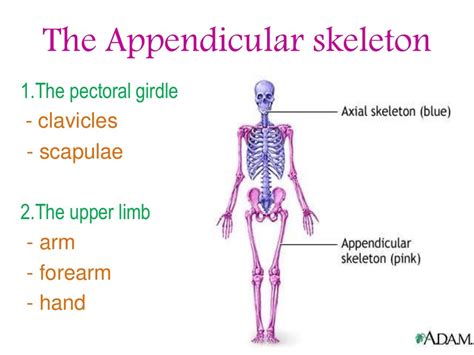
Table of Contents
Which of the Following is Part of the Appendicular Skeleton? A Deep Dive into Human Anatomy
The human skeleton is a marvel of engineering, providing structure, support, and protection for our bodies. It's broadly divided into two main parts: the axial skeleton and the appendicular skeleton. While the axial skeleton forms the central axis of the body (skull, vertebral column, rib cage), the appendicular skeleton comprises the appendages—the limbs and the girdles that attach them to the axial skeleton. Understanding this distinction is crucial for anyone studying anatomy, physiology, or related fields. This article will delve deep into the appendicular skeleton, clarifying which bones belong and exploring their individual functions and significance.
Understanding the Appendicular Skeleton: More Than Just Arms and Legs
The appendicular skeleton isn't simply a collection of bones; it's a complex system of interconnected components working in harmony. It's responsible for locomotion, manipulation of objects, and overall body movement. The key components include:
-
The Pectoral Girdle (Shoulder Girdle): This connects the upper limbs to the axial skeleton. It's composed of the clavicle (collarbone) and the scapula (shoulder blade). Its remarkable flexibility allows for a wide range of arm movements.
-
The Upper Limbs: These are the arms, forearms, and hands. They consist of the humerus (upper arm bone), radius and ulna (forearm bones), carpals (wrist bones), metacarpals (palm bones), and phalanges (finger bones). The intricate structure of the hand allows for precise manipulation and dexterity.
-
The Pelvic Girdle (Hip Girdle): This robust structure connects the lower limbs to the axial skeleton. It's formed by the fusion of three bones: the ilium, ischium, and pubis. The pelvic girdle provides stability and support for the lower body.
-
The Lower Limbs: These are the legs, feet, and thighs. They consist of the femur (thigh bone), patella (kneecap), tibia and fibula (lower leg bones), tarsals (ankle bones), metatarsals (foot bones), and phalanges (toe bones). The strong bones of the lower limbs support our weight and enable locomotion.
Detailed Breakdown of Appendicular Skeleton Bones
Let's break down each component of the appendicular skeleton in more detail, highlighting key features and functions:
1. The Pectoral Girdle: Mobility and Flexibility
The pectoral girdle, unlike the pelvic girdle, is relatively lightly constructed, allowing for a greater degree of mobility.
-
Clavicle (Collarbone): A long, S-shaped bone that connects the sternum (breastbone) to the scapula. It acts as a strut, transferring forces from the upper limb to the axial skeleton. It also helps to keep the shoulder joint stable. Fractures of the clavicle are relatively common, often occurring from falls or direct impacts.
-
Scapula (Shoulder Blade): A flat, triangular bone situated on the back of the rib cage. It articulates with the clavicle and the humerus, forming the shoulder joint. Its unique shape and the associated muscles allow for a wide range of arm movements, including flexion, extension, abduction, adduction, rotation, and circumduction. The scapula's mobility is crucial for activities like throwing, swimming, and lifting objects.
2. The Upper Limbs: Precision and Dexterity
The upper limbs are designed for precision and dexterity.
-
Humerus (Upper Arm Bone): The longest bone in the upper limb. It articulates with the scapula at the shoulder joint and with the radius and ulna at the elbow joint. The humerus plays a crucial role in arm movements, and its proximal end (near the shoulder) contains several important features, including the head, greater and lesser tubercles, and surgical neck.
-
Radius and Ulna (Forearm Bones): These two bones run parallel to each other, allowing for rotation of the forearm (pronation and supination). The radius is the thicker of the two and is located on the lateral side of the forearm. The ulna is located on the medial side and plays a crucial role in elbow stability.
-
Carpals (Wrist Bones): Eight small bones arranged in two rows. These bones allow for a wide range of wrist movements. The carpals form a complex articulation with the radius and ulna, creating a flexible joint.
-
Metacarpals (Palm Bones): Five long bones forming the palm of the hand. These bones connect the carpals to the phalanges.
-
Phalanges (Finger Bones): Fourteen bones in each hand, forming the fingers. The thumb has two phalanges (proximal and distal), while each of the other fingers has three (proximal, middle, and distal). The structure of the phalanges allows for the precise manipulation of objects.
3. The Pelvic Girdle: Stability and Support
The pelvic girdle is a much more stable structure compared to the pectoral girdle.
-
Ilium: The largest bone of the pelvic girdle, forming the upper part of the hip bone. It articulates with the sacrum (part of the vertebral column) forming the sacroiliac joint.
-
Ischium: Forms the lower and back part of the hip bone. It bears weight when seated.
-
Pubis: Forms the anterior (front) part of the hip bone. The two pubic bones meet at the pubic symphysis, a cartilaginous joint.
The fusion of these three bones forms the acetabulum, a deep socket that receives the head of the femur, forming the hip joint. The pelvic girdle's stability is essential for weight-bearing and locomotion. The shape of the female pelvis differs significantly from the male pelvis, reflecting its role in childbirth.
4. The Lower Limbs: Weight-Bearing and Locomotion
The lower limbs are designed for weight-bearing and locomotion.
-
Femur (Thigh Bone): The longest and strongest bone in the body. It articulates with the acetabulum at the hip joint and with the tibia and patella at the knee joint. The femur plays a critical role in weight-bearing and locomotion.
-
Patella (Kneecap): A sesamoid bone (embedded in a tendon) that protects the knee joint and enhances the action of the quadriceps muscle.
-
Tibia and Fibula (Lower Leg Bones): The tibia (shinbone) is the weight-bearing bone of the lower leg, while the fibula provides lateral stability. They articulate with the femur at the knee joint and with the talus (ankle bone) at the ankle joint.
-
Tarsals (Ankle Bones): Seven bones that form the ankle and help support the weight of the body. The talus is the most important tarsal bone, articulating with the tibia and fibula.
-
Metatarsals (Foot Bones): Five long bones forming the arch of the foot. These bones connect the tarsals to the phalanges.
-
Phalanges (Toe Bones): Fourteen bones in each foot, forming the toes. The structure of the phalanges contributes to balance and locomotion.
Common Injuries and Conditions Affecting the Appendicular Skeleton
The appendicular skeleton, due to its role in movement and weight-bearing, is susceptible to several injuries and conditions. These include:
-
Fractures: Broken bones are common, particularly in the clavicle, humerus, radius, ulna, femur, and tibia. These fractures can range from simple hairline cracks to complex comminuted fractures.
-
Dislocations: The shoulder and knee joints are particularly prone to dislocations, where the bones are forced out of their normal alignment.
-
Sprains and Strains: Overstretching or tearing of ligaments (sprain) or muscles and tendons (strain) are common injuries, often occurring in the ankle, knee, wrist, and shoulder.
-
Osteoporosis: A condition characterized by decreased bone density, making bones more fragile and prone to fractures. This condition is more common in older adults, particularly women.
-
Osteoarthritis: A degenerative joint disease causing cartilage breakdown and joint pain. This condition commonly affects the knees, hips, and hands.
Conclusion: The Importance of the Appendicular Skeleton
The appendicular skeleton is a vital part of the human body, responsible for mobility, manipulation, and weight-bearing. Understanding its structure, function, and the potential for injuries is crucial for anyone interested in human anatomy, physiology, or related fields. From the delicate bones of the hand to the strong bones of the legs, each component plays a vital role in our daily lives. The intricate interplay between bones, muscles, and ligaments allows for a remarkable range of movement and dexterity. Taking care of our appendicular skeleton through proper nutrition, exercise, and injury prevention is essential for maintaining overall health and well-being. This detailed exploration should enhance your understanding of this critical skeletal system and its contribution to human function.
Latest Posts
Latest Posts
-
The Number Of Protons In An Atom Is Called
Mar 14, 2025
-
Which Of The Following Cells Is Phagocytic
Mar 14, 2025
-
Meiosis Results In The Production Of
Mar 14, 2025
-
What Is It Called When It Says Speed With Direction
Mar 14, 2025
-
During Aerobic Cellular Respiration The Final Electron Acceptor Is
Mar 14, 2025
Related Post
Thank you for visiting our website which covers about Which Of The Following Is Part Of The Appendicular Skeleton . We hope the information provided has been useful to you. Feel free to contact us if you have any questions or need further assistance. See you next time and don't miss to bookmark.
