What Is The Study Of Tissue Called
News Leon
Mar 30, 2025 · 6 min read
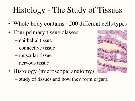
Table of Contents
What is the Study of Tissue Called? An In-Depth Look at Histology
The study of tissues is called histology. This fascinating field bridges the gap between microscopic cellular structures and the macroscopic anatomy and physiology of organisms. Understanding histology is crucial for advancements in numerous fields, from medicine and pathology to botany and zoology. This comprehensive guide will delve deep into the world of histology, exploring its techniques, applications, and significance in various scientific disciplines.
What is a Tissue?
Before diving into the intricacies of histology, it's essential to define its fundamental subject: tissue. A tissue is a group of similar cells and their extracellular matrix (ECM) that work together to perform a specific function. These cells are interconnected and coordinated, forming a cohesive unit within a larger organism. The ECM, a complex network of proteins and carbohydrates, provides structural support and facilitates communication between cells.
The Four Basic Tissue Types
The animal kingdom, and indeed much of multicellular life, organizes itself into four primary tissue types:
1. Epithelial Tissue: The Covering and Lining Expert
Epithelial tissue forms linings of body cavities, organs, and covers the external surfaces. Think of the skin, the lining of your digestive tract, or the cells that make up your lungs. Its key characteristics include:
- Cellularity: Epithelial tissue is composed almost entirely of cells with minimal ECM.
- Specialized Contacts: Cells are tightly bound together through junctions like tight junctions, adherens junctions, desmosomes, and gap junctions. This creates a barrier that protects underlying tissues.
- Polarity: Epithelial cells exhibit apical (free) and basal (attached) surfaces, with distinct structures and functions on each side.
- Support: Epithelial tissue rests on a basement membrane, a thin layer of ECM separating it from underlying connective tissue.
- Avascular: Epithelial tissue lacks blood vessels; it receives nutrients via diffusion from underlying connective tissue.
- Regeneration: Epithelial tissue has a high capacity for regeneration, replacing damaged or worn-out cells.
Types of Epithelial Tissue: Epithelial tissue is further classified based on cell shape (squamous, cuboidal, columnar) and cell arrangement (simple, stratified, pseudostratified). Each type has unique structural and functional adaptations suited to its location and role.
2. Connective Tissue: The Supportive Maestro
Connective tissue is the most abundant and diverse tissue type. Its primary function is to support, connect, and separate different tissues and organs. Key characteristics include:
- Abundant ECM: Connective tissues have a significant amount of ECM, which provides structural support and allows for the diffusion of nutrients and waste products.
- Varied Cell Types: Connective tissue contains a variety of specialized cells, including fibroblasts (responsible for producing ECM), adipocytes (fat cells), chondrocytes (cartilage cells), osteocytes (bone cells), and blood cells.
- Vascularity: Most connective tissues have a rich blood supply, except for cartilage and tendons, which are relatively avascular.
Types of Connective Tissue: The vast range of connective tissue includes loose connective tissue (areolar, adipose, reticular), dense connective tissue (regular, irregular, elastic), cartilage (hyaline, elastic, fibrocartilage), bone, and blood. Each type has distinct properties and functions.
3. Muscle Tissue: The Movement Specialist
Muscle tissue is responsible for movement, both voluntary and involuntary. Its key feature is the presence of contractile proteins (actin and myosin) that allow for muscle cell shortening and force generation.
- Skeletal Muscle: Attached to bones, responsible for voluntary movement. Characterized by long, cylindrical, multinucleated cells with striations (alternating light and dark bands).
- Smooth Muscle: Found in the walls of internal organs and blood vessels. Responsible for involuntary movements, like digestion and blood pressure regulation. Characterized by spindle-shaped, uninucleated cells lacking striations.
- Cardiac Muscle: Found exclusively in the heart. Responsible for the rhythmic contractions of the heart. Characterized by branched, uninucleated cells with intercalated discs (specialized junctions that allow for rapid communication between cells).
4. Nervous Tissue: The Communication Network
Nervous tissue forms the central and peripheral nervous systems. It is specialized for rapid communication and coordination of body functions. Its key features are:
- Neurons: Specialized cells that transmit electrical signals (nerve impulses) throughout the body. Neurons have a cell body (soma), dendrites (receiving signals), and an axon (transmitting signals).
- Neuroglia: Supporting cells that provide structural and metabolic support for neurons. Examples include astrocytes, oligodendrocytes, and microglia.
Histological Techniques: Unveiling the Microscopic World
Histological techniques are crucial for preparing and visualizing tissues under a microscope. These techniques involve several steps:
- Fixation: Preserving the tissue's structure and preventing degradation.
- Processing: Removing water and embedding the tissue in a solid medium (e.g., paraffin wax) for sectioning.
- Sectioning: Cutting thin slices (sections) of the tissue using a microtome.
- Staining: Applying dyes to highlight specific cellular components or structures. Common stains include hematoxylin and eosin (H&E), which stain nuclei and cytoplasm respectively. Specialized stains can be used to visualize specific components such as collagen, elastin, or lipids.
- Mounting: Placing the stained sections onto microscope slides for observation.
Applications of Histology
Histology is an indispensable tool across numerous scientific disciplines:
- Pathology: Diagnosing diseases by examining tissue samples (biopsies). Cancer diagnosis heavily relies on histological examination.
- Medical Research: Investigating the effects of diseases, drugs, and environmental factors on tissues.
- Forensic Science: Identifying tissue samples in criminal investigations.
- Veterinary Medicine: Diagnosing and treating diseases in animals.
- Botany: Studying the structure and function of plant tissues.
- Zoology: Investigating the tissues of various animal species.
Histopathology: The Intersection of Histology and Pathology
Histopathology is a specialized branch of histology focusing on the microscopic examination of diseased tissues. It's crucial for accurate disease diagnosis, particularly in cancer. Histopathologists analyze tissue samples to identify abnormal cells, assess the extent of tissue damage, and determine the stage and grade of a disease. This information is essential for guiding treatment strategies and predicting prognosis.
Advanced Histological Techniques
Beyond traditional techniques, advancements in technology have led to several sophisticated methods:
- Immunohistochemistry (IHC): Using antibodies to detect specific proteins in tissues, aiding in the diagnosis of various diseases, including cancers.
- In situ hybridization (ISH): Detecting specific nucleic acid sequences (DNA or RNA) in tissues, allowing for the localization of genes and their expression patterns.
- Electron Microscopy: Utilizing electron beams to visualize ultra-fine structures within cells and tissues, providing higher resolution than light microscopy.
- Confocal Microscopy: Using lasers to create sharp images of thick tissue sections, allowing for 3D reconstruction of tissues.
The Future of Histology
The field of histology is constantly evolving. New techniques and technologies are continuously being developed, pushing the boundaries of what we can observe and understand about tissue structure and function. Advances in imaging, data analysis, and artificial intelligence are shaping the future of histology, enabling more precise diagnoses, personalized medicine, and a deeper understanding of biological processes.
Conclusion: The Essential Role of Histology
Histology, the study of tissues, is a fundamental discipline with far-reaching applications. From diagnosing diseases to advancing medical research, histology provides critical insights into the structure and function of living organisms. Its sophisticated techniques continue to evolve, promising an even deeper understanding of the complex world of tissues and their significance in health and disease. The crucial role histology plays in numerous fields underscores its importance as a cornerstone of biological and medical science. The ongoing advancements in techniques and technology promise to further enhance our ability to utilize histology's power in diagnosis, treatment, and research, ultimately improving human and animal health.
Latest Posts
Latest Posts
-
Greatest Amount Of Digestion Takes Place In The
Apr 01, 2025
-
All Real Numbers Are Rational Numbers True Or False
Apr 01, 2025
-
Which Of The Following Is A Function That Money Serves
Apr 01, 2025
-
The Most Abundant Compound In Most Living Things Is
Apr 01, 2025
-
How Can We Change The Polarity Of An Electromagnet
Apr 01, 2025
Related Post
Thank you for visiting our website which covers about What Is The Study Of Tissue Called . We hope the information provided has been useful to you. Feel free to contact us if you have any questions or need further assistance. See you next time and don't miss to bookmark.
