What Is The Structure Indicated By Label E
News Leon
Mar 15, 2025 · 5 min read
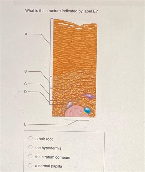
Table of Contents
What is the Structure Indicated by Label E? A Deep Dive into Biological Structures
The question "What is the structure indicated by label E?" is a common one in biology, particularly in histology and anatomy. Without knowing the specific image or diagram in question, a precise answer is impossible. However, we can explore various potential structures that might be labeled "E" depending on the context. This comprehensive guide will delve into several possibilities, providing detailed explanations and relevant examples. We'll explore the structural components, functions, and clinical significance of these structures, ensuring a thorough understanding of what "E" might represent.
Potential Structures Labeled "E": A Comprehensive Overview
The label "E" could refer to a vast array of biological structures, depending on the accompanying diagram or micrograph. To provide the most helpful response, we'll explore several potential candidates, categorized for clarity.
1. Cellular Structures:
-
Eukaryotic Cell Organelles: If the diagram depicts a cell, "E" could signify a specific organelle. Possibilities include:
-
Endoplasmic Reticulum (ER): The ER is a network of membranes involved in protein synthesis and lipid metabolism. The rough ER (RER), studded with ribosomes, is responsible for protein synthesis, while the smooth ER (SER) plays a role in lipid synthesis and detoxification. A diagram might label the ER as "E," indicating its extensive network within the cell.
-
Golgi Apparatus (Golgi Body): This organelle processes and packages proteins and lipids for secretion or delivery to other cellular compartments. Its characteristic stacked structure would make it easily identifiable.
-
Mitochondria: These are the "powerhouses" of the cell, responsible for cellular respiration and ATP production. Their distinctive double-membrane structure, with cristae within the inner membrane, would be a key identifying feature.
-
Lysosomes: These membrane-bound organelles contain hydrolytic enzymes that break down waste materials and cellular debris. Their identification depends on their size and location within the cell.
-
Nucleolus: Located within the nucleus, the nucleolus is the site of ribosome synthesis. It appears as a densely stained region within the nucleus.
-
-
Prokaryotic Cell Structures: In diagrams of prokaryotic cells (bacteria and archaea), "E" could represent:
-
Ribosomes: These are essential for protein synthesis in both prokaryotic and eukaryotic cells. They are smaller in prokaryotes than eukaryotes.
-
Plasmid: These are small, circular DNA molecules found in many bacteria, often carrying genes that provide advantages such as antibiotic resistance.
-
Capsule: A protective layer surrounding some bacteria, composed of polysaccharides or polypeptides.
-
2. Tissue Structures:
If the diagram represents a tissue sample, "E" could indicate a variety of structural elements.
-
Connective Tissue Components: In connective tissue, "E" might indicate:
-
Extracellular Matrix (ECM): This is the non-cellular component of connective tissue, composed of ground substance and fibers (collagen, elastin, reticulin). The ECM provides structural support and mediates cell-cell interactions.
-
Specific Fiber Types: The diagram might highlight specific collagen fibers or elastin fibers, indicating their orientation and distribution within the tissue.
-
Cells within Connective Tissue: "E" could also point to specific cell types like fibroblasts (responsible for collagen production), chondrocytes (cartilage cells), or osteocytes (bone cells).
-
-
Epithelial Tissue Components: In epithelial tissue, "E" could label:
-
Cell Junctions: These are specialized structures that connect epithelial cells, creating a cohesive layer. Examples include tight junctions, adherens junctions, desmosomes, and gap junctions.
-
Basement Membrane: This is a thin layer of extracellular matrix that separates the epithelium from underlying connective tissue.
-
-
Muscle Tissue Components: In muscle tissue, "E" might refer to:
-
Sarcomeres: The basic contractile unit of muscle fibers, composed of actin and myosin filaments.
-
Myofibrils: Long, cylindrical structures within muscle fibers, composed of numerous sarcomeres.
-
3. Organ Structures:
At the organ level, "E" might label:
-
Specific Layers or Regions: In complex organs like the heart or kidney, "E" could indicate a specific layer (e.g., the myocardium of the heart or the renal medulla of the kidney).
-
Blood Vessels: "E" might identify specific blood vessels within an organ, such as arteries, veins, or capillaries.
-
Nerve Bundles or Ganglia: In organs with significant nervous innervation, "E" could label nerve bundles or ganglia.
The Importance of Context: Deciphering the Label
The critical element in determining the structure indicated by "E" is the context provided by the accompanying diagram or micrograph. Consider the following:
-
Magnification: A high-magnification image might show cellular structures, while a lower-magnification image might depict tissues or organs.
-
Labels and Captions: Pay close attention to any labels or captions that might offer clues about the tissue type or organ being depicted.
-
Staining Techniques: Different staining techniques highlight specific cellular or tissue components. Knowing the staining technique used can provide valuable insights into the structure's identity.
-
Overall Structure: The overall architecture of the structure, including its shape, size, and relationship to other structures, is crucial for identification.
Clinical Significance: Why Understanding Structure Matters
Understanding the structure indicated by "E," regardless of its identity, has significant clinical implications. Accurate identification is critical for:
-
Diagnosis: Many diseases manifest as changes in tissue structure. Identifying the affected structure is the first step towards accurate diagnosis.
-
Treatment Planning: The choice of treatment often depends on the specific structure involved. For example, treatment for a disease affecting the liver will differ from treatment for a disease affecting the kidney.
-
Prognosis: The structure's condition and response to treatment can provide insights into the prognosis of a disease.
-
Research: Understanding the structure and function of different biological structures is crucial for advancing our understanding of health and disease.
Conclusion: A Multifaceted Approach
Determining the structure indicated by label "E" requires a systematic and multifaceted approach. By carefully considering the context, magnification, staining techniques, and overall structure, along with a solid understanding of various biological structures, we can confidently identify the component in question. Remember, accuracy in identification is crucial for effective diagnosis, treatment, and furthering our understanding of the complex world of biology. This necessitates not just memorization but a deeper understanding of the interrelationships between different biological structures and their functional significance within the organism. The more we understand, the better equipped we are to tackle the complexities of biological study and clinical practice.
Latest Posts
Latest Posts
-
Is Osmosis High To Low Or Low To High
Mar 15, 2025
-
Concave Mirror And Convex Mirror Difference
Mar 15, 2025
-
Which Is Not A Cranial Bone Of The Skull
Mar 15, 2025
-
Mountain Range That Separates Europe And Asia
Mar 15, 2025
-
16 Out Of 40 As A Percentage
Mar 15, 2025
Related Post
Thank you for visiting our website which covers about What Is The Structure Indicated By Label E . We hope the information provided has been useful to you. Feel free to contact us if you have any questions or need further assistance. See you next time and don't miss to bookmark.
