What Is The Purpose Of Simple Staining
News Leon
Apr 05, 2025 · 6 min read
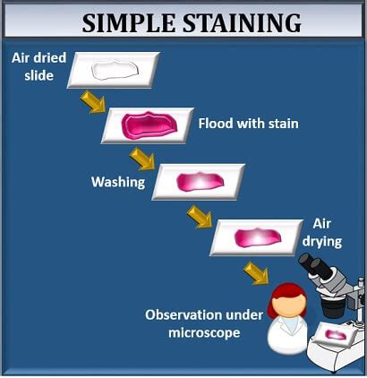
Table of Contents
What is the Purpose of Simple Staining? A Comprehensive Guide
Simple staining, a fundamental technique in microbiology, serves a crucial purpose: to visualize the basic morphology of microorganisms. While seemingly straightforward, its applications extend far beyond a simple observation. Understanding the purpose of simple staining requires delving into its methodology, applications, and limitations. This comprehensive guide will explore these aspects in detail, equipping you with a thorough understanding of this essential microbiological technique.
Understanding the Mechanics of Simple Staining
Simple staining employs a single basic dye to color bacterial cells, rendering them visible against a contrasting background. The process relies on the differential charge between the bacterial cell and the dye. Bacterial cells typically possess a negatively charged surface due to the presence of teichoic acids in Gram-positive bacteria and lipopolysaccharides in Gram-negative bacteria. Basic dyes, such as methylene blue, crystal violet, and safranin, are positively charged, thus forming an electrostatic attraction with the negatively charged bacterial surface. This interaction leads to the staining of the cells, making them easily observable under a light microscope.
Key Components of Simple Staining:
-
Basic Dye: The core element, providing the color to the bacterial cells. The choice of dye can influence the intensity and shade of staining, but the primary purpose remains consistent. Popular choices include:
- Methylene blue: A commonly used dye known for its versatility and relatively low toxicity.
- Crystal violet: Produces a darker, more intense stain, making it suitable for visualizing certain cell structures.
- Safranin: A counterstain often used in Gram staining but also applicable in simple staining, offering a reddish-pink hue.
-
Microscope Slide: A clean, grease-free slide is essential to ensure proper adherence of the bacterial smear.
-
Microscope: A compound light microscope is necessary to visualize the stained bacteria. The magnification needed will depend on the size of the microorganisms and the level of detail required.
-
Bacterial Smear: A thin, even layer of bacterial cells spread on the slide, crucial for accurate observation and avoiding overlapping cells that obscure the morphology.
The Simple Staining Procedure:
The simple staining procedure is relatively straightforward, typically involving the following steps:
- Smear Preparation: A small amount of bacterial culture is spread thinly and evenly on a clean microscope slide. This is allowed to air dry completely.
- Heat Fixation: The dried smear is gently passed over a Bunsen burner flame to fix the cells to the slide. This process kills the bacteria and prevents them from being washed away during staining. Overheating must be avoided to prevent cell distortion.
- Dye Application: The chosen basic dye is applied to the smear for a specific duration, typically 1-2 minutes.
- Rinsing: Excess dye is gently rinsed off using distilled water.
- Blot Drying: The slide is carefully blotted dry with bibulous paper.
- Microscopic Examination: The stained smear is examined under a light microscope at appropriate magnification.
The Primary Purpose: Visualizing Bacterial Morphology
The most fundamental purpose of simple staining is to reveal the shape and arrangement of bacterial cells. By staining the cells, the background becomes transparent, making the microorganisms easily distinguishable. This allows microbiologists to identify key morphological characteristics, including:
- Cocci (spherical): These can appear singly, in pairs (diplococci), in chains (streptococci), in clusters (staphylococci), or in tetrads (groups of four).
- Bacilli (rod-shaped): These can be single, in pairs (diplobacilli), in chains (streptobacilli), or arranged in palisades (side-by-side).
- Spiral/Helical: These include spirilla (rigid spirals) and spirochetes (flexible spirals).
Observing these morphological features is the first step in bacterial identification. While simple staining doesn't provide detailed information about cell structure, it provides a crucial foundation for further, more sophisticated analyses.
Beyond Basic Morphology: Extended Applications of Simple Staining
While primarily used for visualizing basic morphology, simple staining finds applications in other areas:
-
Quick Assessment of Bacterial Viability: While not a precise measure, a simple stain can offer a quick estimate of bacterial viability. A high number of stained cells indicates a likely high viability, although this method doesn't distinguish between live and dead cells accurately.
-
Initial Screening for Contamination: In various settings, from food microbiology to clinical laboratories, simple staining can be a rapid method for assessing the presence of bacterial contamination.
-
Educational Tool: Simple staining is an excellent tool for teaching basic microbiological techniques. Its simplicity allows students to grasp fundamental principles of microscopy and staining before progressing to more complex techniques.
-
Preliminary Step for Other Staining Techniques: In some cases, simple staining can act as a preliminary step for more complex staining procedures, such as Gram staining or acid-fast staining. It allows for a quick assessment of the sample before undertaking more involved staining protocols.
Limitations of Simple Staining
It's crucial to acknowledge the limitations of simple staining:
-
Lack of Differential Staining: Simple staining doesn't differentiate between different types of bacteria. It stains all bacterial cells similarly, unlike differential stains (like Gram staining) that differentiate based on cell wall characteristics.
-
Limited Information on Cell Structure: Simple staining only reveals basic morphology. It doesn't provide information about internal cell structures, such as flagella, capsules, or endospores. Specialized staining techniques are required to visualize these structures.
-
Potential for Artifacts: Improper smear preparation or heat fixation can introduce artifacts, potentially leading to misinterpretations of bacterial morphology.
-
Overlapping Cells: A thick smear can lead to overlapping cells, obscuring individual cell shapes and making accurate observation challenging.
Simple Staining vs. Differential Staining: A Comparison
Simple staining's limitations become apparent when compared to differential staining techniques, such as Gram staining and acid-fast staining. While simple staining provides a general overview of morphology, differential staining provides further insights based on specific characteristics:
| Feature | Simple Staining | Differential Staining (e.g., Gram stain) |
|---|---|---|
| Number of dyes | One | Two or more |
| Purpose | Visualize basic morphology | Differentiate bacteria based on cell wall properties |
| Information | Shape, arrangement | Shape, arrangement, cell wall type |
| Complexity | Simple, quick | More complex, multi-step |
| Applications | Initial screening, education | Bacterial identification, diagnosis |
Conclusion: The Enduring Value of Simple Staining
Despite its limitations, simple staining remains an indispensable tool in microbiology. Its simplicity, speed, and effectiveness in visualizing basic bacterial morphology make it invaluable for a variety of applications. From basic educational purposes to quick assessments of microbial populations, simple staining provides a fundamental starting point for understanding the microbial world. While it may not provide the level of detail offered by more sophisticated techniques, its role in foundational microbiological practices remains crucial and enduring. Understanding the purpose and limitations of simple staining is essential for any microbiologist, regardless of their area of specialization.
Latest Posts
Latest Posts
-
Differences Between Onion Epidermal And Human Epithelial Cells
Apr 05, 2025
-
Choose The True Statement About The Krebs Cycle
Apr 05, 2025
-
Five More Than The Quotient Of A Number And 4
Apr 05, 2025
-
Hollow Spherical Shell Moment Of Inertia
Apr 05, 2025
-
Which Of The Following Is True For Displacement
Apr 05, 2025
Related Post
Thank you for visiting our website which covers about What Is The Purpose Of Simple Staining . We hope the information provided has been useful to you. Feel free to contact us if you have any questions or need further assistance. See you next time and don't miss to bookmark.
