The Space Between The Cornea And The Iris Is The
News Leon
Mar 21, 2025 · 6 min read
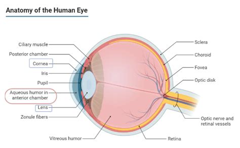
Table of Contents
The Space Between the Cornea and the Iris Is the Anterior Chamber: A Deep Dive into Anatomy, Function, and Clinical Significance
The human eye, a marvel of biological engineering, is a complex organ responsible for our sense of sight. Understanding its intricate structure is crucial to appreciating its functionality and the potential implications of various pathologies. One key component often overlooked is the anterior chamber, the space situated between the cornea and the iris. This article delves deep into the anatomy, physiology, and clinical significance of this vital structure.
Anatomy of the Anterior Chamber: A Microscopic View
The anterior chamber is a fluid-filled space, shaped like a wedge, occupying the area between the cornea anteriorly and the iris and lens posteriorly. Its boundaries are precisely defined:
-
Anteriorly: The endothelium of the cornea forms the anterior boundary. This single layer of cells plays a crucial role in maintaining corneal transparency and hydration. Damage to the corneal endothelium can severely compromise vision.
-
Posteriorly: The iris, with its intricate network of blood vessels and pigment cells, forms the posterior boundary. The iris's pupil, a central opening, regulates the amount of light entering the eye.
-
Peripherally: The anterior chamber angle, a crucial area, is formed by the junction of the cornea and the iris. This angle is where the trabecular meshwork resides, a spongy network of tissues responsible for draining aqueous humor from the eye. The angle's structure is critical for maintaining intraocular pressure (IOP).
The anterior chamber is not merely an empty space; it's filled with aqueous humor, a clear, watery fluid produced by the ciliary body. This fluid nourishes the cornea and lens, maintaining their transparency and health. The constant production and drainage of aqueous humor is vital for regulating intraocular pressure, preventing damage to the delicate structures of the eye.
The Significance of the Anterior Chamber Angle
The anterior chamber angle's structure is incredibly significant. Its openness or narrowness directly affects the outflow of aqueous humor. A narrow angle can lead to angle-closure glaucoma, a serious condition characterized by a sudden increase in intraocular pressure due to blocked outflow of aqueous humor. This blockage can cause severe damage to the optic nerve, potentially leading to irreversible vision loss. Conversely, an open angle allows for normal aqueous humor drainage.
Physiology of the Anterior Chamber: Maintaining Intraocular Pressure
The anterior chamber plays a crucial role in maintaining the eye's intraocular pressure (IOP). This pressure is the result of a delicate balance between the production and drainage of aqueous humor. The ciliary body, located behind the iris, continuously produces aqueous humor, which flows into the posterior chamber and then through the pupil into the anterior chamber. From there, the aqueous humor drains primarily through the trabecular meshwork in the anterior chamber angle, ultimately entering the Schlemm's canal and then the venous system.
Any disruption in this delicate balance can lead to alterations in IOP. Increased IOP, as seen in glaucoma, can cause damage to the optic nerve and lead to vision loss. Conversely, decreased IOP can also be problematic, potentially indicating underlying conditions.
The Role of Aqueous Humor in Nourishment and Transparency
Aqueous humor is not merely involved in pressure regulation; it also plays a vital role in nourishing the avascular cornea and lens. These structures lack blood vessels and rely on the nutrients and oxygen supplied by the aqueous humor for their metabolic needs. The composition of aqueous humor is carefully controlled, ensuring the proper balance of nutrients and waste products.
The clarity of aqueous humor is essential for maintaining visual acuity. Any clouding or inflammation of the aqueous humor can significantly impair vision.
Clinical Significance of the Anterior Chamber: Common Conditions and Diagnostic Procedures
The anterior chamber's structure and function are closely linked to several clinically significant conditions:
1. Glaucoma: The Silent Thief of Sight
Glaucoma, a group of eye diseases, is characterized by increased intraocular pressure (IOP). This elevated pressure damages the optic nerve, potentially leading to irreversible vision loss. Angle-closure glaucoma is directly related to the anatomy of the anterior chamber angle. Narrow angles can obstruct the outflow of aqueous humor, leading to a sudden and dramatic increase in IOP. Open-angle glaucoma, a more common form, is characterized by a gradual increase in IOP due to impaired drainage through the trabecular meshwork, even though the angle itself remains open.
2. Iritis and Uveitis: Inflammation of the Iris and Uvea
Iritis, inflammation of the iris, and uveitis, inflammation of the uvea (the middle layer of the eye containing the iris, ciliary body, and choroid), can significantly affect the anterior chamber. Inflammation can cause the formation of inflammatory cells and proteins in the aqueous humor, leading to clouding of the anterior chamber and impaired vision. These conditions often require prompt medical intervention to prevent irreversible damage.
3. Hyphema: Blood in the Anterior Chamber
Hyphema, the presence of blood in the anterior chamber, is a serious condition often caused by trauma to the eye. Blood can obstruct vision and potentially lead to further complications if not treated promptly. The accumulation of blood in the anterior chamber can increase IOP and cause damage to the corneal endothelium.
4. Anterior Chamber Depth Measurement: A Key Diagnostic Tool
Measuring the anterior chamber depth (ACD) is a crucial diagnostic procedure, often used in the evaluation of glaucoma risk. A shallow anterior chamber is a risk factor for angle-closure glaucoma. ACD measurement can be performed using various techniques, including ultrasound biomicroscopy and optical coherence tomography.
5. Gonioscopy: Visualizing the Anterior Chamber Angle
Gonioscopy is a specialized examination technique used to visualize the anterior chamber angle. This procedure is essential for evaluating the openness of the angle and identifying any abnormalities that may contribute to glaucoma or other conditions. Gonioscopy uses a special lens to allow the examiner to view the angle structures directly.
Advanced Imaging Techniques: A Deeper Look Inside
Modern imaging techniques provide detailed visualization of the anterior chamber and its surrounding structures, enhancing diagnostic capabilities:
-
Ultrasound Biomicroscopy (UBM): UBM provides high-resolution images of the anterior segment of the eye, including the anterior chamber, iris, and angle structures. It's particularly useful in evaluating angle anatomy and identifying abnormalities associated with glaucoma.
-
Anterior Segment Optical Coherence Tomography (AS-OCT): AS-OCT offers high-resolution, cross-sectional imaging of the anterior segment. It's valuable for assessing the thickness of the cornea, the depth of the anterior chamber, and the morphology of the angle structures.
-
Confocal Microscopy: This advanced technique allows for microscopic visualization of living tissues, providing detailed images of the cellular components of the anterior chamber and corneal endothelium.
Conclusion: The Anterior Chamber – A Vital Component of Ocular Health
The anterior chamber, though seemingly a small and simple space, plays a multifaceted and crucial role in maintaining ocular health. Its anatomical structure, the dynamic interplay of aqueous humor production and drainage, and its susceptibility to various pathologies underscore its importance. Understanding the anterior chamber's anatomy, physiology, and clinical significance is paramount for ophthalmologists and other healthcare professionals involved in the diagnosis and management of eye diseases. The advancement of imaging techniques continues to refine our ability to visualize and understand this vital component of the eye, leading to improved diagnostic accuracy and treatment strategies. Early detection and intervention are key to preventing irreversible vision loss associated with conditions affecting the anterior chamber. Regular comprehensive eye examinations are essential for maintaining optimal ocular health and preventing the development or progression of conditions that affect this critical structure.
Latest Posts
Latest Posts
-
Elements And Compounds Are Two Types Of
Mar 21, 2025
-
Definition Of Average Acceleration In Physics
Mar 21, 2025
-
Opportunity Cost Occurs Because Of A Producers Need To
Mar 21, 2025
-
How Many Chambers Does A Frogs Heart Have
Mar 21, 2025
-
Is Burning Of Paper A Chemical Change
Mar 21, 2025
Related Post
Thank you for visiting our website which covers about The Space Between The Cornea And The Iris Is The . We hope the information provided has been useful to you. Feel free to contact us if you have any questions or need further assistance. See you next time and don't miss to bookmark.
