The Serous Membrane That Covers The Lungs Is The
News Leon
Mar 16, 2025 · 6 min read
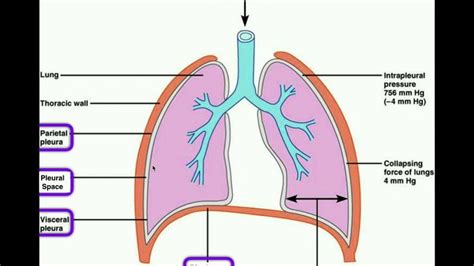
Table of Contents
The Serous Membrane That Covers the Lungs Is the Pleura: A Deep Dive into Pulmonary Anatomy and Physiology
The lungs, the vital organs responsible for gas exchange, are not simply floating freely within the thoracic cavity. They are enveloped and protected by a thin, slippery serous membrane known as the pleura. Understanding the pleura's structure, function, and clinical significance is crucial for comprehending respiratory physiology and various pulmonary pathologies. This comprehensive article will delve into the intricacies of the pleura, exploring its anatomy, its role in lung mechanics, and the implications of pleural disorders.
Anatomy of the Pleura: A Double-Layered System
The pleura is a delicate, double-layered serous membrane that completely encloses each lung. It consists of two continuous but distinct layers:
1. Visceral Pleura: The Lung's Inner Lining
The visceral pleura is the innermost layer, intimately adherent to the surface of each lung, including its fissures and extending into the lung hilum. It's essentially a part of the lung itself, following every contour and indentation. Its smooth surface minimizes friction during lung expansion and contraction. The visceral pleura is richly innervated, providing sensory information about the lungs and the surrounding structures.
2. Parietal Pleura: The Thoracic Cavity's Inner Lining
The parietal pleura lines the inner surface of the thoracic cavity, adhering to the chest wall, diaphragm, mediastinum, and superior thoracic aperture. Unlike the visceral pleura, it is less directly associated with lung tissue and is innervated by somatic nerves, rendering it highly sensitive to pain. This layer has several distinct parts, based on its anatomical location:
- Costal pleura: Covers the inner surface of the ribs and intercostal muscles.
- Diaphragmatic pleura: Covers the superior surface of the diaphragm.
- Mediastinal pleura: Lines the mediastinum, the central compartment of the thorax containing the heart, great vessels, and esophagus.
- Cervical pleura (cupula): Extends superiorly into the neck, reaching the root of the neck.
The Pleural Cavity: A Potential Space
Between the visceral and parietal pleura lies the pleural cavity, a potential space containing a small amount of serous fluid. This fluid, known as pleural fluid, acts as a lubricant, reducing friction between the two pleural layers during respiration. The extremely low pressure within the pleural cavity (subatmospheric pressure) is critical for maintaining lung inflation.
Physiological Role of the Pleura in Respiration: A Crucial Partnership
The pleura's contribution to respiratory mechanics is paramount. Its structural integrity and the unique properties of the pleural fluid are indispensable for normal breathing.
1. Lung Expansion and Contraction: The Pleural Link
During inspiration, the diaphragm contracts and flattens, and the intercostal muscles elevate the ribs. This action expands the thoracic cavity, increasing its volume. Because of the negative pressure within the pleural cavity, the lungs are passively pulled outwards, expanding along with the chest wall. This cohesive linkage between the parietal and visceral pleura, mediated by the pleural fluid, prevents the lungs from collapsing.
During expiration, the diaphragm and intercostal muscles relax, decreasing the volume of the thoracic cavity. The elastic recoil of the lungs, along with the surface tension of the alveolar fluid, helps to expel air from the lungs. The pleural pressure increases slightly, but remains negative throughout the respiratory cycle, maintaining lung expansion.
2. Surface Tension and Lung Compliance: The Pleural Fluid's Role
Pleural fluid significantly contributes to lung compliance, the ease with which the lungs expand. The surface tension of the pleural fluid, along with the elastic properties of lung tissue, influence how much effort is required for inspiration and expiration. Any disruption to the normal composition or volume of pleural fluid can affect lung compliance, leading to respiratory distress.
Clinical Significance of Pleural Disorders: A Wide Spectrum of Diseases
Pleural disorders encompass a wide range of conditions affecting the pleura, significantly impacting respiratory function. Some common examples include:
1. Pleuritis (Pleurisy): Inflammation of the Pleura
Pleuritis, or pleurisy, is characterized by inflammation of the pleural membranes. It's often associated with underlying conditions like pneumonia, lung cancer, tuberculosis, or autoimmune diseases. The hallmark symptom is sharp, stabbing chest pain, particularly during breathing or coughing. Inflammation can cause the pleural surfaces to rub against each other, creating a characteristic pleural friction rub, audible with a stethoscope.
2. Pleural Effusion: Fluid Accumulation in the Pleural Cavity
A pleural effusion involves an abnormal accumulation of fluid in the pleural cavity. This fluid can be transudative (due to increased hydrostatic pressure or decreased oncotic pressure) or exudative (due to inflammation or infection). The presence of a pleural effusion can compress the lung, reducing its ability to expand and impair gas exchange. Symptoms vary depending on the amount and type of fluid, ranging from shortness of breath to chest pain.
3. Pneumothorax: Air in the Pleural Cavity
A pneumothorax is the presence of air in the pleural cavity, leading to lung collapse. It can occur spontaneously (due to a bleb or bulla rupture), traumatically (due to chest injury), or iatrogenically (due to medical procedures). A pneumothorax can cause sudden, sharp chest pain and shortness of breath. In severe cases, it can be life-threatening, requiring immediate medical attention.
4. Mesothelioma: A Rare and Aggressive Cancer
Mesothelioma is a rare and aggressive cancer arising from the mesothelial cells lining the pleura. Exposure to asbestos is the primary risk factor. Symptoms often develop late in the disease, making early diagnosis challenging. Treatment options are limited, and the prognosis is often poor.
5. Pleural Thickening: Scarring and Fibrosis of the Pleura
Pleural thickening is characterized by scarring and fibrosis of the pleural membranes, often due to previous inflammation or infection. It can restrict lung expansion and impair respiratory function. The condition may be asymptomatic or lead to shortness of breath, particularly during exertion.
Diagnostic Approaches to Pleural Disorders: Essential Tools for Assessment
Several diagnostic tools are used to assess pleural disorders:
- Chest X-ray: A standard imaging technique to detect pleural effusions, pneumothorax, and pleural thickening.
- Computed Tomography (CT) scan: Provides detailed cross-sectional images of the chest, allowing for better visualization of pleural abnormalities.
- Ultrasound: Useful for guiding thoracentesis (fluid removal from the pleural cavity) and evaluating the characteristics of pleural fluid.
- Thoracentesis: A procedure to remove pleural fluid for analysis to determine its composition and identify the underlying cause.
- Pleural biopsy: A procedure to obtain a tissue sample from the pleura for histological examination.
Conclusion: The Pleura's Significance in Respiratory Health
The pleura, a seemingly inconspicuous serous membrane, plays a pivotal role in maintaining respiratory function. Its unique anatomical structure and physiological properties are essential for lung expansion and contraction, gas exchange, and overall respiratory health. Understanding the anatomy, physiology, and clinical significance of the pleura is vital for healthcare professionals involved in diagnosing and managing respiratory conditions. A multitude of disorders can affect the pleura, ranging from inflammation and fluid accumulation to cancer, underscoring the importance of early diagnosis and effective treatment strategies for improving patient outcomes and ensuring respiratory well-being. Further research continues to unravel the intricacies of pleural biology and to develop novel therapeutic approaches for pleural disorders.
Latest Posts
Latest Posts
-
A Charge Of Uniform Linear Density
Mar 17, 2025
-
Which Of The Following Is Polynomial
Mar 17, 2025
-
Is Boiling Water A Physical Change
Mar 17, 2025
-
Is A Webcam An Input Or Output Device
Mar 17, 2025
-
Word For A Person Who Uses Big Words
Mar 17, 2025
Related Post
Thank you for visiting our website which covers about The Serous Membrane That Covers The Lungs Is The . We hope the information provided has been useful to you. Feel free to contact us if you have any questions or need further assistance. See you next time and don't miss to bookmark.
