The Inspiratory And Expiratory Centers Are Located In The
News Leon
Mar 21, 2025 · 6 min read
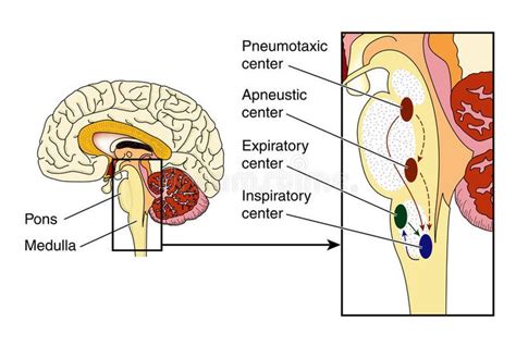
Table of Contents
The Inspiratory and Expiratory Centers: Location, Function, and Clinical Significance
The rhythmic process of breathing, essential for life, is orchestrated by a complex network of neural circuits residing primarily within the brainstem. Central to this control are the inspiratory and expiratory centers, which regulate the timing and depth of inhalation and exhalation. Understanding their precise location, intricate interplay, and clinical implications is crucial for grasping the intricacies of respiratory physiology and associated pathologies.
Location of the Respiratory Centers
While the term "centers" might suggest discrete, anatomically defined nuclei, the respiratory control system is more accurately described as a network of interconnected neuronal populations distributed across several brainstem regions. The primary location for these control mechanisms lies within the medulla oblongata and pons, two crucial components of the brainstem.
Medulla Oblongata: The Primary Respiratory Center
The medulla oblongata houses the two key groups of neurons directly involved in the basic rhythm of breathing:
-
Dorsal Respiratory Group (DRG): Located in the dorsal part of the medulla, the DRG is predominantly responsible for inhalation. It receives sensory input from peripheral chemoreceptors (detecting changes in blood oxygen and carbon dioxide levels) and mechanoreceptors in the lungs (monitoring lung stretch). The DRG primarily activates the phrenic nerve, which innervates the diaphragm, the primary muscle of inspiration, and also influences the intercostal nerves, which control the intercostal muscles involved in rib cage expansion. The DRG generates the basic rhythm of breathing, although its activity is modulated by other centers.
-
Ventral Respiratory Group (VRG): Situated in the ventral medulla, the VRG plays a more complex role, becoming increasingly active during increased respiratory demands. While the DRG predominantly controls inspiration, the VRG is involved in both inspiration and expiration. During normal quiet breathing, the VRG's activity is minimal. However, during forceful breathing (e.g., exercise, hyperventilation), the VRG's expiratory neurons become active, facilitating active exhalation by stimulating expiratory muscles. The VRG also contains pre-Botzinger complex, a group of neurons believed to be crucial for generating the rhythmic pattern of breathing.
Pons: Modulatory Influence on Respiratory Rhythm
The pons, located superior to the medulla, doesn't generate the basic respiratory rhythm but plays a significant modulatory role, refining and modifying the output from the medullary centers:
-
Pneumotaxic Center: This center, located in the upper pons, primarily functions to limit inspiration. It sends inhibitory signals to the DRG, preventing overinflation of the lungs. The pneumotaxic center's activity helps regulate the respiratory rate and the depth of breathing. It acts as a "switch" which turns off inspiration. A high frequency of signals from this center results in shorter and shallower breaths, whereas lower frequencies lead to slower and deeper breaths.
-
Apneustic Center: Situated in the lower pons, the apneustic center has the opposite effect of the pneumotaxic center. It promotes inspiration, prolonging the inspiratory phase. The exact function of the apneustic center is less clear than the pneumotaxic center, and its role might depend on the interaction with other respiratory centers. Lesions affecting this area can lead to prolonged inspirations, a condition known as apneusis.
Integration and Control: A Complex Interplay
The respiratory centers don't function in isolation. Instead, they interact in a complex, integrated manner to fine-tune the respiratory response to various physiological demands. This intricate interplay involves several mechanisms:
-
Feedback Loops: Sensory feedback from peripheral chemoreceptors, lung stretch receptors, and other mechanoreceptors constantly provides information about the respiratory system's status. This feedback is integrated by the respiratory centers to adjust the breathing pattern accordingly. For example, an increase in blood carbon dioxide levels triggers increased ventilation to expel the excess CO2.
-
Higher Brain Centers: While the brainstem controls the basic rhythm, higher brain centers, such as the hypothalamus and cerebral cortex, can exert significant influence on respiration. For instance, emotional responses (fear, anxiety) or conscious control (holding one's breath) can override the brainstem's autonomic control.
-
Chemical Factors: Blood gases (oxygen, carbon dioxide) and pH play critical roles in regulating respiration. Chemoreceptors sensitive to these factors provide input to the respiratory centers, modulating breathing according to the body's metabolic needs.
-
Neurotransmitters: Various neurotransmitters, including glutamate, GABA, and serotonin, mediate communication within and between the respiratory centers. The balance of these neurotransmitters profoundly impacts respiratory rhythm and pattern.
Clinical Significance: Respiratory Disorders and Dysfunction
Dysfunction in any part of this intricate network can lead to various respiratory disorders. Understanding the location and function of the respiratory centers is essential for diagnosing and managing these conditions:
-
Central Sleep Apnea: This condition is characterized by repeated pauses in breathing during sleep due to dysfunction in the respiratory centers' ability to maintain appropriate ventilation.
-
Ondine's Curse (Congenital Central Hypoventilation Syndrome): This rare disorder is caused by a genetic defect affecting the development or function of the respiratory centers. Affected individuals require artificial ventilation, as their brainstem cannot automatically control breathing.
-
Brainstem Stroke: Damage to the medulla or pons can severely impair respiration, leading to respiratory arrest or irregular breathing patterns. The location of the stroke within the brainstem often determines the type of respiratory dysfunction observed.
-
Medullary Depression: Certain drugs or toxins can depress the activity of the medullary respiratory centers, resulting in slowed or inadequate breathing. This is a life-threatening condition requiring immediate medical attention.
-
Respiratory Failure: This can result from various causes, including lung disease, neuromuscular disorders, or dysfunction of the respiratory centers. Respiratory failure necessitates artificial ventilation to support breathing.
-
Neurodegenerative Diseases: Conditions like amyotrophic lateral sclerosis (ALS) can affect the motor neurons involved in respiratory control, gradually leading to respiratory muscle weakness and failure.
-
Post-Polio Syndrome: Years after acute poliomyelitis infection, some individuals may experience new respiratory symptoms due to continued degeneration of motor neurons.
Investigating Respiratory Control: Diagnostic Tools
Neurological assessment, including observation of respiratory patterns (rate, depth, rhythm), is crucial in diagnosing respiratory dysfunction. Further investigations may include:
-
Polysomnography: This sleep study is valuable in diagnosing sleep apnea by monitoring breathing patterns, heart rate, and brain activity during sleep.
-
Electroencephalography (EEG): EEG can help assess brainwave patterns and identify potential abnormalities in the brainstem's respiratory control regions.
-
Magnetic Resonance Imaging (MRI): MRI can provide high-resolution images of the brainstem, allowing for the detection of structural abnormalities that might affect respiratory function.
-
Blood gas analysis: Measurement of blood oxygen and carbon dioxide levels provides crucial information about respiratory efficiency.
Conclusion: A Vital System Requiring Precise Regulation
The inspiratory and expiratory centers, distributed across the medulla oblongata and pons, orchestrate the essential process of breathing. Their complex interactions, modulated by various internal and external factors, ensure a finely tuned respiratory response to physiological demands. Understanding the location and function of these centers is fundamental in comprehending normal respiratory physiology and the pathogenesis of numerous respiratory disorders. Further research into the intricate neural circuitry and molecular mechanisms governing respiratory control will likely lead to improved diagnostic tools and therapeutic strategies for respiratory diseases. The continuous interplay between the medullary and pontine regions, with crucial modulation from higher brain centers and peripheral feedback loops, underscores the complexity and vital importance of this system for sustaining life. Appreciating this intricacy allows for a deeper understanding of the delicate balance required for efficient respiration and the potential consequences of disruptions within this essential network.
Latest Posts
Latest Posts
-
In The Figure A Metal Wire Of Mass
Mar 21, 2025
-
Find Three Consecutive Even Integers Whose Sum Is 108
Mar 21, 2025
-
A Catalyst Lowers The Activation Energy Of A Reaction By
Mar 21, 2025
-
Formic Acid And Sodium Hydroxide Balanced Equation
Mar 21, 2025
-
Where In A Plant Cell Is Chlorophyll Found
Mar 21, 2025
Related Post
Thank you for visiting our website which covers about The Inspiratory And Expiratory Centers Are Located In The . We hope the information provided has been useful to you. Feel free to contact us if you have any questions or need further assistance. See you next time and don't miss to bookmark.
