The Contractile Unit Of A Myofibril Is Called The
News Leon
Mar 22, 2025 · 6 min read
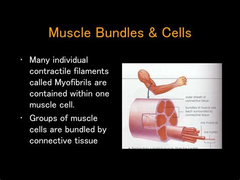
Table of Contents
The Contractile Unit of a Myofibril is Called the Sarcomere: A Deep Dive into Muscle Contraction
The human body is a marvel of engineering, capable of feats of strength, endurance, and precision. At the heart of this capability lies the muscle, a tissue composed of specialized cells that contract to generate movement. Understanding how muscles work requires delving into their intricate structure, down to the smallest functional unit. This article will explore the sarcomere, the contractile unit of a myofibril, and delve into the fascinating mechanisms that power muscle contraction.
What is a Myofibril?
Before we dive into the sarcomere, let's establish the context. Skeletal muscles, the muscles responsible for voluntary movement, are composed of bundles of muscle fibers. Each muscle fiber is a single, multinucleated cell containing numerous cylindrical structures called myofibrils. These myofibrils are the fundamental units responsible for the muscle's contractile properties. They are packed with highly organized protein filaments arranged in a repeating pattern, giving them a characteristic striated appearance under a microscope. This striated pattern is directly linked to the structure and function of the sarcomere.
The Sarcomere: The Basic Contractile Unit
The sarcomere is the fundamental unit of striated muscle tissue. It's the smallest functional unit capable of contraction. Think of it as the tiny engine driving muscle movement. Each sarcomere is a highly organized structure extending from one Z-line (or Z-disc) to the next Z-line. These Z-lines are protein structures that serve as anchoring points for the contractile filaments.
Key Components of a Sarcomere:
-
Z-lines (Z-discs): These are the boundaries of the sarcomere. They are dense protein structures that anchor the thin filaments (actin).
-
A-band (Anisotropic band): This is the dark band in the striated pattern. It represents the entire length of the thick filaments (myosin), including the regions where thick and thin filaments overlap.
-
I-band (Isotropic band): This is the light band in the striated pattern. It contains only thin filaments (actin) and extends from the A-band of one sarcomere to the A-band of the adjacent sarcomere. The Z-line bisects the I-band.
-
H-zone: Located in the center of the A-band, this is a lighter region within the A-band containing only thick filaments (myosin). It disappears during maximal muscle contraction.
-
M-line: This is a dark line found in the center of the H-zone. It's a structural protein that anchors the thick filaments (myosin) and plays a crucial role in maintaining sarcomere organization.
-
Thick Filaments (Myosin): These are composed primarily of the protein myosin, a motor protein with a head and tail region. The myosin heads interact with the thin filaments to generate force during contraction.
-
Thin Filaments (Actin): These are composed primarily of the protein actin, along with other regulatory proteins like tropomyosin and troponin. Tropomyosin blocks myosin binding sites on actin in a resting muscle, while troponin plays a key role in calcium-mediated regulation of muscle contraction.
The Sliding Filament Theory: How Sarcomeres Contract
The mechanism by which sarcomeres shorten and generate force is explained by the sliding filament theory. This theory postulates that muscle contraction occurs as a result of the overlapping thick and thin filaments sliding past each other without changing their individual lengths.
Steps in Muscle Contraction:
-
Neural Stimulation: Muscle contraction begins with a nerve impulse reaching the neuromuscular junction. This triggers the release of acetylcholine, a neurotransmitter that depolarizes the muscle fiber's membrane.
-
Calcium Release: Depolarization initiates a cascade of events that lead to the release of calcium ions (Ca2+) from the sarcoplasmic reticulum, a specialized intracellular calcium store.
-
Calcium Binding to Troponin: The released Ca2+ binds to troponin, a protein complex located on the thin filaments. This binding causes a conformational change in troponin, which moves tropomyosin away from the myosin-binding sites on actin.
-
Cross-Bridge Formation: With the myosin-binding sites on actin now exposed, the myosin heads can bind to actin, forming cross-bridges.
-
Power Stroke: After binding, the myosin head undergoes a conformational change, pivoting and pulling the thin filament towards the center of the sarcomere. This is the power stroke, generating the force of muscle contraction.
-
Cross-Bridge Detachment: ATP binds to the myosin head, causing it to detach from actin.
-
ATP Hydrolysis and Myosin Reset: ATP is hydrolyzed (broken down) into ADP and inorganic phosphate (Pi). This provides the energy for the myosin head to return to its original conformation, ready to bind to another actin molecule and repeat the cycle.
-
Sarcomere Shortening: The repeated cycle of cross-bridge formation, power stroke, detachment, and resetting leads to the sliding of thin filaments over thick filaments, resulting in sarcomere shortening and overall muscle contraction.
-
Calcium Removal: Once the nerve impulse ceases, calcium ions are actively pumped back into the sarcoplasmic reticulum, removing them from troponin. This causes tropomyosin to return to its blocking position, preventing further cross-bridge formation and muscle relaxation.
Types of Muscle Contractions:
Understanding sarcomere function allows us to understand the different types of muscle contractions:
-
Isometric Contraction: Muscle tension increases, but the muscle length remains the same. This occurs when the force generated is not sufficient to overcome the load. Think of holding a heavy object in place.
-
Isotonic Contraction: Muscle tension remains constant, but the muscle length changes. This is further divided into:
- Concentric Contraction: Muscle shortens while generating force (e.g., lifting a weight).
- Eccentric Contraction: Muscle lengthens while generating force (e.g., lowering a weight slowly).
Factors Affecting Sarcomere Function and Muscle Contraction:
Several factors influence the efficiency and strength of muscle contraction:
-
Number of Sarcomeres: Muscles with a higher number of sarcomeres in parallel can generate greater force.
-
Sarcomere Length: Optimal sarcomere length is essential for maximal force generation. Too short or too long a sarcomere will reduce the number of cross-bridges that can form.
-
ATP Availability: ATP is essential for both the power stroke and cross-bridge detachment. ATP depletion leads to muscle fatigue.
-
Calcium Ion Concentration: Adequate calcium ion concentration is crucial for initiating and maintaining muscle contraction.
-
Neural Input: The frequency and strength of neural stimulation influence the rate and strength of muscle contraction.
Sarcomere Dysfunction and Muscle Diseases:
Disruptions in sarcomere structure and function are implicated in various muscle diseases, including:
-
Muscular Dystrophies: These are a group of genetic disorders characterized by progressive muscle weakness and degeneration. Mutations in genes encoding proteins involved in sarcomere structure can lead to instability and dysfunction.
-
Myotonic Dystrophy: This is a group of inherited disorders affecting muscle relaxation. It’s caused by mutations that disrupt the function of ion channels involved in muscle excitation and contraction.
-
Heart Failure: Sarcomere dysfunction plays a crucial role in heart failure, where impaired contractility leads to reduced pumping efficiency.
Conclusion:
The sarcomere, the contractile unit of a myofibril, is a remarkably intricate and efficient structure. Its precise organization and the sophisticated mechanisms of the sliding filament theory allow for the generation of force and movement essential for life. Understanding the sarcomere's structure and function is critical for comprehending muscle physiology, diagnosing muscle diseases, and developing therapeutic interventions. Further research into sarcomere dynamics promises to unlock even greater insights into the complexities of muscle function and human movement. This detailed understanding helps us appreciate the incredible power and precision of our own bodies, from the smallest sarcomere to the largest muscle group. The ongoing study of sarcomeres continues to unveil new discoveries in the field of biomechanics and muscle physiology, offering exciting potential for future advancements in medicine and rehabilitation.
Latest Posts
Latest Posts
-
Complete The Following Table Regarding Acids And Bases
Mar 22, 2025
-
Which Of The Following Statements About Dna Replication Is True
Mar 22, 2025
-
X 3 2x 2 5x 10
Mar 22, 2025
-
Which Of These Nuclides Is Most Likely To Be Radioactive
Mar 22, 2025
-
What Is The Conjugate Base Of Hpo42
Mar 22, 2025
Related Post
Thank you for visiting our website which covers about The Contractile Unit Of A Myofibril Is Called The . We hope the information provided has been useful to you. Feel free to contact us if you have any questions or need further assistance. See you next time and don't miss to bookmark.
