Respiratory Control Centers Are Located In The ________.
News Leon
Mar 28, 2025 · 6 min read
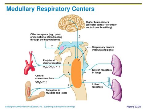
Table of Contents
Respiratory Control Centers are Located in the Brainstem
The regulation of breathing, a fundamental process for survival, is intricately controlled by a network of neurons clustered in the brainstem. This isn't a single, centralized location, but rather a complex interplay between several respiratory centers located within the medulla oblongata and pons. Understanding the precise locations and functions of these centers is crucial to comprehending how we breathe, both at rest and during physical activity, and how disruptions in these areas can lead to respiratory disorders.
The Medulla Oblongata: The Primary Respiratory Control Center
The medulla oblongata, the lower part of the brainstem, houses two crucial respiratory centers: the dorsal respiratory group (DRG) and the ventral respiratory group (VRG). These groups work in concert, coordinating the rhythm and depth of our breathing.
Dorsal Respiratory Group (DRG): Setting the Basic Rhythm
The DRG is considered the primary pacemaker for breathing. Located in the nucleus tractus solitarius (NTS), it receives sensory input from various peripheral chemoreceptors and mechanoreceptors. These receptors monitor blood gas levels (oxygen, carbon dioxide) and lung stretch, providing crucial feedback to the DRG.
-
Inspiration: The DRG primarily controls inspiration, the process of inhaling. Neurons within the DRG fire rhythmically, activating inspiratory muscles like the diaphragm and external intercostal muscles. This rhythmic firing creates the basic rhythm of breathing. The duration and intensity of these signals determine the depth and rate of breathing.
-
Sensory Input: The DRG's function is profoundly influenced by sensory input. For example, increased carbon dioxide levels (hypercapnia) or decreased oxygen levels (hypoxemia) detected by peripheral chemoreceptors stimulate the DRG to increase the rate and depth of breathing. Similarly, stretch receptors in the lungs (pulmonary stretch receptors) send signals to the DRG, preventing overinflation of the lungs through the Hering-Breuer reflex.
Ventral Respiratory Group (VRG): Fine-Tuning Breathing
While the DRG sets the basic rhythm, the VRG plays a crucial role in fine-tuning the respiratory pattern, particularly during increased respiratory demand like exercise. Located in the medulla's ventrolateral region, the VRG contains several neuronal populations that contribute to both inspiration and expiration.
-
Inspiration and Expiration: Unlike the DRG, the VRG's activity isn't solely restricted to inspiration. It contains neurons that actively participate in both inspiration and expiration. This allows for the recruitment of accessory muscles during strenuous activity, increasing the force and volume of both inhalation and exhalation. These neurons can fine-tune the rhythm and depth of breathing in response to changing metabolic demands.
-
Pre-Bötzinger Complex: A critical part of the VRG is the pre-Bötzinger complex (pre-BötC), which plays a vital role in generating the respiratory rhythm. Research strongly suggests that the pre-BötC is the primary rhythm generator, creating the basic respiratory pattern that the DRG and VRG then modify. The exact mechanisms underlying the pre-BötC’s rhythm-generating properties are still being investigated, but it involves complex neuronal interactions and intricate network properties.
The Pons: Modifying and Fine-Tuning Medullary Output
The pons, located superior to the medulla, contains two important respiratory centers: the pneumotaxic center and the apneustic center. These centers don't generate the basic respiratory rhythm but significantly influence the output from the medullary centers, modifying the rate and depth of breathing.
Pneumotaxic Center: Limiting Inspiration
The pneumotaxic center primarily functions to limit inspiration. It sends signals to the DRG, shortening the duration of inspiratory bursts. This results in a faster, shallower breathing pattern. The pneumotaxic center's influence is crucial for regulating the transition between inspiration and expiration, ensuring a smooth and coordinated respiratory cycle. Its activity is inversely proportional to the duration of inspiration; increased pneumotaxic activity leads to shorter inspirations and a higher respiratory rate.
Apneustic Center: Promoting Inspiration
In contrast to the pneumotaxic center, the apneustic center promotes prolonged inspiration. Its signals to the DRG increase the duration of inspiratory bursts, leading to slower, deeper breaths. The apneustic center's activity is counterbalanced by the pneumotaxic center. The interplay between these two centers allows for precise control over respiratory timing and depth. Damage to these centers can lead to significant disruptions in respiratory patterns, such as apneusis (prolonged inspiratory gasps).
Chemical Control of Respiration: Chemoreceptors and Blood Gas Levels
The respiratory centers are highly sensitive to changes in blood gas levels. Chemoreceptors, specialized cells that detect changes in the chemical composition of blood, play a crucial role in this chemical control of respiration. There are two main types of chemoreceptors:
-
Central Chemoreceptors: Located in the medulla, central chemoreceptors are highly sensitive to changes in the cerebrospinal fluid's (CSF) carbon dioxide concentration. Carbon dioxide readily crosses the blood-brain barrier, and an increase in blood CO2 leads to increased CO2 in the CSF. This, in turn, increases the acidity (decreases pH) of the CSF, stimulating central chemoreceptors. This stimulation sends signals to the respiratory centers, increasing breathing rate and depth to eliminate the excess CO2.
-
Peripheral Chemoreceptors: Located in the carotid bodies (at the bifurcation of the common carotid arteries) and aortic bodies (in the aortic arch), peripheral chemoreceptors are sensitive to changes in both blood oxygen and carbon dioxide levels, as well as blood pH. A decrease in blood oxygen (hypoxemia), an increase in blood carbon dioxide (hypercapnia), or a decrease in blood pH (acidosis) stimulates these chemoreceptors. They send signals to the respiratory centers via cranial nerves (glossopharyngeal and vagus nerves), resulting in increased respiratory drive.
Other Influences on Respiration
While the brainstem respiratory centers are primarily responsible for breathing control, other brain regions and factors can influence respiratory patterns:
-
Higher brain centers: The cerebral cortex and limbic system can voluntarily influence breathing, allowing for conscious control over breath-holding and changes in breathing patterns during emotional responses.
-
Mechanoreceptors: Stretch receptors in the lungs and airways monitor lung volume and prevent overinflation. Irritant receptors in the airways trigger coughing and sneezing in response to foreign particles or irritants.
-
Temperature: Increased body temperature can stimulate respiration, while decreased body temperature has the opposite effect.
-
Hormones: Several hormones can influence respiration, particularly during stress or illness.
Clinical Significance: Respiratory Disorders
Disruptions in the function of the brainstem respiratory centers or their inputs can lead to various respiratory disorders, including:
-
Central apnea: Characterized by periods of absent breathing due to dysfunction in the respiratory centers.
-
Ondine's curse (congenital central hypoventilation syndrome): A rare genetic disorder affecting the respiratory centers, resulting in inadequate breathing during sleep.
-
Cheyne-Stokes respiration: A cyclical pattern of breathing characterized by periods of apnea followed by increasing depth of breathing, often seen in patients with heart failure or brain injury.
Conclusion: A Complex and Vital System
The respiratory control centers located in the brainstem form a complex and tightly regulated system essential for life. The interplay between the medullary and pontine respiratory centers, the influence of chemoreceptors, and the input from various other sources ensures finely tuned control of breathing, adapting to both resting conditions and changing metabolic demands. Understanding the intricacies of this system is not only critical for appreciating the normal physiology of breathing but also for diagnosing and managing a range of respiratory disorders. Further research into the precise mechanisms and interactions within this network continues to unravel the complexities of respiratory control. The precise location of the respiratory centers within the medulla and pons underscores the delicate balance required to maintain a life-sustaining process. Continued research continues to illuminate the intricate details of this vital system.
Latest Posts
Latest Posts
-
What Two Factors Does Air Pressure Depend On
Mar 31, 2025
-
Which Lewis Electron Dot Diagram Is Correct For Co2
Mar 31, 2025
-
Which Statement Is True About Obligate Anaerobes
Mar 31, 2025
-
How Many Lines Of Symmetry Circle
Mar 31, 2025
-
Are Chloroplast Found In Animal Cells
Mar 31, 2025
Related Post
Thank you for visiting our website which covers about Respiratory Control Centers Are Located In The ________. . We hope the information provided has been useful to you. Feel free to contact us if you have any questions or need further assistance. See you next time and don't miss to bookmark.
