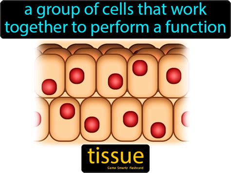
Groups Of Cells Sharing Similar Morphology And Function Form Tissue.
News Leon
Apr 03, 2025 · 6 min read

Table of Contents
Groups of Cells Sharing Similar Morphology and Function Form Tissue: A Deep Dive into Histology
The human body, a marvel of biological engineering, is not a monolithic structure. Instead, it's a complex hierarchy of organization, starting with the fundamental building blocks: cells. These cells, while diverse in their specialized roles, often group together to form tissues—collections of cells with similar morphology (structure) and function. Understanding tissues is crucial to comprehending the workings of organs, organ systems, and ultimately, the entire organism. This article will explore the fascinating world of tissues, examining their classification, properties, and significance in maintaining overall health.
The Foundation of Life: Cells and Their Organization
Before diving into the complexities of tissues, let's briefly revisit the concept of cells. Cells are the basic units of life, each carrying out essential functions necessary for survival. However, a single cell rarely acts in isolation. To perform more complex tasks, cells cooperate, forming structured communities that enhance their collective efficiency. This cooperation is the essence of tissue formation. Cells with similar characteristics and functions aggregate, establishing communication networks and coordinating their activities to achieve a common purpose.
Key Cellular Characteristics Contributing to Tissue Formation
Several cellular attributes play a crucial role in tissue development and maintenance:
-
Cell Adhesion Molecules (CAMs): These specialized molecules on cell surfaces facilitate cell-to-cell binding, crucial for maintaining tissue integrity and structure. Different CAMs mediate various types of cell junctions, contributing to the unique properties of different tissues.
-
Extracellular Matrix (ECM): This intricate network of proteins and polysaccharides surrounds cells, providing structural support, regulating cell behavior, and influencing tissue properties. The composition and organization of the ECM vary significantly across different tissue types, reflecting their unique functional demands.
-
Cell Signaling: Cells within a tissue constantly communicate through chemical signals, coordinating their activities and maintaining tissue homeostasis. This communication is crucial for growth, repair, and overall tissue function. Disruptions in cell signaling can lead to various pathologies.
-
Cell Differentiation: The process by which a cell becomes specialized for a specific function is critical for tissue development. Stem cells, with their capacity for self-renewal and differentiation, give rise to diverse cell types within a tissue, ensuring its functionality.
The Four Primary Tissue Types: An Overview
Histologists, scientists who study tissues, typically classify tissues into four primary types based on their structure and function:
- Epithelial Tissue: Covers body surfaces, lines body cavities and organs, and forms glands.
- Connective Tissue: Supports, connects, and separates different tissues and organs.
- Muscle Tissue: Enables movement through contraction.
- Nervous Tissue: Transmits electrical signals throughout the body, enabling communication and coordination.
Epithelial Tissue: The Protective Shield
Epithelial tissue is characterized by closely packed cells with minimal extracellular matrix. It forms continuous sheets that cover body surfaces, line body cavities and organs, and constitute the functional units of glands. Its primary functions include protection, secretion, absorption, excretion, filtration, diffusion, and sensory reception. The unique arrangement and specialization of epithelial cells dictate their specific roles.
Classification of Epithelial Tissue:
Epithelial tissues are classified based on two key features:
- Cell Shape: Squamous (flat), cuboidal (cube-shaped), and columnar (tall and column-shaped).
- Number of Cell Layers: Simple (single layer) and stratified (multiple layers).
Examples:
- Simple Squamous Epithelium: Found in the lining of blood vessels (endothelium) and body cavities (mesothelium), facilitating diffusion and filtration.
- Stratified Squamous Epithelium: Forms the epidermis of the skin, providing protection against abrasion and dehydration.
- Simple Cuboidal Epithelium: Lines ducts and tubules of glands, involved in secretion and absorption.
- Stratified Cuboidal Epithelium: Relatively rare, found in some ducts of glands.
- Simple Columnar Epithelium: Lines the digestive tract, facilitating absorption and secretion. Often contains goblet cells, which secrete mucus.
- Stratified Columnar Epithelium: Found in certain ducts and parts of the male urethra.
- Pseudostratified Columnar Epithelium: Appears stratified but is actually a single layer of cells, found in the respiratory tract, often ciliated to move mucus.
- Transitional Epithelium: Lines the urinary tract, capable of stretching and changing shape to accommodate changes in urine volume.
Connective Tissue: The Body's Support System
Connective tissues are characterized by abundant extracellular matrix, which separates widely spaced cells. This matrix, composed of ground substance and fibers (collagen, elastic, reticular), provides structural support, binds tissues together, and facilitates communication between cells. Connective tissues exhibit remarkable diversity in their structure and function, reflecting their varied roles throughout the body.
Types of Connective Tissue:
-
Connective Tissue Proper: Includes loose connective tissue (areolar, adipose, reticular) and dense connective tissue (regular, irregular, elastic). Loose connective tissue provides support and cushioning, while dense connective tissue offers strength and resistance to stress.
-
Specialized Connective Tissue: Includes cartilage (hyaline, elastic, fibrocartilage), bone, and blood. Cartilage provides flexible support, bone provides rigid support and protection, and blood transports oxygen and nutrients.
Examples:
- Adipose Tissue: Stores energy as fat, provides insulation, and cushions organs.
- Bone Tissue: Provides structural support, protection, and mineral storage.
- Cartilage Tissue: Provides flexible support and cushioning in joints.
- Blood Tissue: Transports oxygen, nutrients, hormones, and waste products.
Muscle Tissue: The Engine of Movement
Muscle tissue is specialized for contraction, enabling movement of the body and its internal organs. Three main types of muscle tissue exist:
- Skeletal Muscle: Voluntary, striated muscle attached to bones, responsible for body movement.
- Smooth Muscle: Involuntary, non-striated muscle found in the walls of internal organs and blood vessels, regulating organ function.
- Cardiac Muscle: Involuntary, striated muscle found only in the heart, responsible for pumping blood.
Nervous Tissue: The Communication Network
Nervous tissue is composed of neurons and glial cells. Neurons are specialized cells that transmit electrical signals throughout the body, enabling rapid communication and coordination. Glial cells support and protect neurons. Nervous tissue forms the brain, spinal cord, and nerves, coordinating all bodily functions.
Tissue Repair and Regeneration
Tissue injury triggers a complex repair process involving inflammation, cell proliferation, and tissue remodeling. The capacity for tissue repair and regeneration varies among different tissue types. Epithelial tissues generally regenerate readily, while connective tissues exhibit varying regenerative capabilities. Muscle tissues have limited regenerative capacity, and nervous tissue regeneration is typically restricted. Understanding the mechanisms of tissue repair is crucial for developing effective therapies for tissue injury and disease.
Clinical Significance of Tissue Understanding
A thorough understanding of tissues is paramount in many medical fields. Pathologists examine tissue samples to diagnose diseases, while surgeons must understand tissue properties to perform procedures effectively. Many diseases directly affect tissues, such as cancers (uncontrolled tissue growth), inflammatory diseases (tissue damage due to inflammation), and degenerative diseases (tissue deterioration). Therefore, knowledge of tissue structure, function, and pathology is crucial for effective diagnosis and treatment.
Conclusion
The study of tissues, or histology, offers a fundamental understanding of the intricate organization and function of the human body. The four primary tissue types—epithelial, connective, muscle, and nervous—each exhibit remarkable diversity in structure and function, reflecting their specialized roles in maintaining overall health. Understanding the properties, interactions, and repair mechanisms of these tissues is essential for advancing medical knowledge and improving healthcare. As research continues to unravel the complexities of tissue biology, we can expect further breakthroughs in disease diagnosis, treatment, and regenerative medicine. Further exploration into specific tissue types and their associated pathologies will continue to reveal insights into the remarkable intricacies of the human body.
Latest Posts
Latest Posts
-
Which Of The Following Is A Non Renewable Source Of Energy
Apr 04, 2025
-
What Binds To The Exposed Cross Bridges On Actin
Apr 04, 2025
-
All Squares Are Rectangles And Rhombuses
Apr 04, 2025
-
What Is The Role Of Toothpaste In Preventing Cavities
Apr 04, 2025
-
Is A Grasshopper A Producer Consumer Or Decomposer
Apr 04, 2025
Related Post
Thank you for visiting our website which covers about Groups Of Cells Sharing Similar Morphology And Function Form Tissue. . We hope the information provided has been useful to you. Feel free to contact us if you have any questions or need further assistance. See you next time and don't miss to bookmark.
