Diagram Of A Simple Reflex Arc
News Leon
Mar 29, 2025 · 7 min read
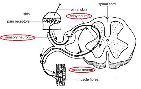
Table of Contents
Diagram of a Simple Reflex Arc: A Deep Dive into Neural Pathways
The human body is a marvel of intricate biological mechanisms, and a prime example of this complexity lies within our nervous system. At the heart of our rapid, involuntary responses to stimuli lies the reflex arc, a neural pathway that mediates reflexes. Understanding the diagram of a simple reflex arc is key to grasping fundamental neurological processes. This comprehensive guide delves into the structure, function, and significance of this fascinating neural pathway.
What is a Reflex Arc?
A reflex arc is a neural pathway that controls a reflex action. A reflex action is an involuntary and nearly instantaneous movement in response to a stimulus. Unlike voluntary actions that involve conscious thought and processing in the brain, reflexes bypass higher brain centers for speed and efficiency. This rapid response is crucial for survival, protecting us from harm by initiating immediate protective actions.
Components of a Simple Reflex Arc: The Diagram Explained
A typical simple reflex arc involves five key components, beautifully illustrated in a diagram:
-
Receptor: This is the specialized sensory nerve ending that detects the stimulus. Different receptors are sensitive to different stimuli, such as pressure, temperature, light, or chemicals. For example, in the knee-jerk reflex, the receptor is a stretch receptor within the muscle spindle of the quadriceps muscle.
-
Sensory Neuron (Afferent Neuron): The receptor triggers the sensory neuron, transmitting the impulse towards the central nervous system (CNS). The sensory neuron's axon carries the signal along its length. The sensory neuron’s cell body is located in the dorsal root ganglion of the spinal cord. This is a crucial point in the diagram; it clearly shows the direction of impulse travel.
-
Interneuron (Association Neuron): In many reflexes, but not all, a connecting neuron, the interneuron, relays the signal between the sensory and motor neurons within the CNS. This neuron is located within the grey matter of the spinal cord. The interneuron plays a crucial role in integrating information and coordinating the response. Its presence is critical in more complex reflexes. Simple reflexes can sometimes bypass the interneuron, establishing a direct connection between the sensory and motor neurons (monosynaptic reflex).
-
Motor Neuron (Efferent Neuron): This neuron carries the impulse from the CNS to the effector organ. The cell body of the motor neuron is located in the anterior horn of the spinal cord, and its axon extends to the muscle. The diagram explicitly shows the motor neuron's role in transmitting the impulse away from the CNS.
-
Effector: This is the muscle or gland that carries out the reflex response. In the case of the knee-jerk reflex, the effector is the quadriceps muscle, which contracts in response to the stimulus. Other effectors include glands, which may secrete hormones or other substances in response to a reflex.
Types of Reflex Arcs
Reflex arcs are not all created equal. They are categorized based on the number of synapses involved:
-
Monosynaptic Reflex Arc: This is the simplest type of reflex arc, involving only one synapse between the sensory neuron and the motor neuron. The knee-jerk reflex is a classic example. The diagram of a monosynaptic reflex arc is particularly straightforward, showcasing the direct connection between the sensory and motor neurons.
-
Polysynaptic Reflex Arc: This type of reflex arc involves two or more synapses, including one or more interneurons. This allows for more complex processing and integration of information. The withdrawal reflex (e.g., pulling your hand away from a hot stove) is a polysynaptic reflex, involving interneurons that coordinate the contraction of flexor muscles and relaxation of extensor muscles. The diagram of a polysynaptic reflex arc is more complex, demonstrating the multiple connections and pathways involved.
Understanding the distinction between these two types helps appreciate the complexity and variability within reflex arcs.
The Importance of the Reflex Arc Diagram in Understanding Neurological Function
A well-drawn diagram is an invaluable tool for understanding the reflex arc. The visual representation clarifies the sequential steps involved in the reflex pathway, emphasizing the direction of impulse transmission and the roles of each component. The diagram provides a clear and concise summary of a complex biological process, making it easier to grasp the key concepts.
Common Examples of Reflexes and their Corresponding Diagrams
Several everyday reflexes exemplify the principles discussed above:
-
Knee-jerk reflex (Patellar reflex): A tap below the patella stretches the quadriceps muscle, activating stretch receptors. This triggers a monosynaptic reflex, resulting in quadriceps contraction and extension of the leg. The diagram shows a direct connection between the sensory and motor neuron.
-
Withdrawal reflex: Touching a hot object stimulates nociceptors (pain receptors) in the skin. This triggers a polysynaptic reflex, causing flexion of the affected limb and withdrawal from the stimulus. The diagram highlights the involvement of interneurons coordinating the muscle contractions.
-
Pupillary light reflex: Shining a bright light into the eye causes the pupil to constrict. This reflex involves sensory neurons detecting light intensity, interneurons in the brainstem, and motor neurons innervating the iris muscles. The diagram shows the complex pathway and the involvement of the brainstem.
Clinical Significance of Reflex Testing
Reflex testing is a crucial component of neurological examinations. By evaluating reflexes, clinicians can assess the integrity of the nervous system. Abnormal reflexes can indicate damage to the nervous system at various levels, including spinal cord injuries, nerve damage, or neurological disorders. The understanding of the reflex arc diagram and its components helps interpret the results of these tests. The absence or exaggeration of a reflex, or the presence of abnormal reflexes, can provide valuable diagnostic information.
Further Exploration of Reflex Arc Mechanisms
Beyond the basic components, several intricate mechanisms modulate reflex arc activity:
-
Synaptic transmission: The transmission of nerve impulses across synapses is a crucial step in the reflex arc. Neurotransmitters play a central role in this process, and their actions are susceptible to modulation. Drugs and toxins can influence synaptic transmission, affecting reflex responses.
-
Reciprocal innervation: In many reflexes, such as the withdrawal reflex, coordinated muscle actions are involved. For example, the contraction of flexor muscles is accompanied by the relaxation of extensor muscles. This reciprocal innervation is facilitated by interneurons within the reflex arc.
-
Central pattern generators: Some reflexes involve rhythmic, repetitive movements, such as walking or breathing. These rhythmic patterns are generated by neural circuits called central pattern generators (CPGs), located within the spinal cord or brainstem. The diagram of a reflex arc is a simplified representation, not showing the complexities of CPGs.
The Role of Feedback Mechanisms in Reflex Control
Reflexes are not simply hardwired, automatic responses. Feedback mechanisms play a crucial role in adjusting and refining reflex activity. For example, sensory feedback from the muscle itself can modulate the strength and duration of the reflex response. This feedback helps ensure that the reflex is appropriate for the specific situation and avoids over- or under-compensation. This dynamic aspect of reflexes is often not captured in simplified diagrams but is crucial for understanding the complete picture.
Conclusion: The Reflex Arc – A Foundation of Neurological Function
The simple reflex arc, though seemingly simple at first glance, represents a fundamental building block of neurological function. Understanding the diagram of a simple reflex arc, its components, and its variations provides insight into the rapid, involuntary responses that protect us from harm and facilitate crucial physiological processes. From the monosynaptic knee-jerk reflex to the more complex polysynaptic withdrawal reflex, the principles of the reflex arc are universally applicable throughout the nervous system. Through a thorough understanding of the reflex arc, a solid foundation is established for further exploration of the intricacies of the human nervous system. The ability to interpret reflex arc diagrams and appreciate their clinical significance is invaluable for anyone interested in biology, neuroscience, or medicine. The elegant simplicity of the reflex arc, coupled with its profound importance, makes it a truly fascinating topic of study.
Latest Posts
Latest Posts
-
Domain And Range For Y 1 X
Apr 01, 2025
-
What Is The Mass Of A Beta Particle
Apr 01, 2025
-
12 Of 150 Is What Number
Apr 01, 2025
-
Draw The Organic Products Formed In The Following Reaction
Apr 01, 2025
-
All The Elements In The Same Period Have The Same
Apr 01, 2025
Related Post
Thank you for visiting our website which covers about Diagram Of A Simple Reflex Arc . We hope the information provided has been useful to you. Feel free to contact us if you have any questions or need further assistance. See you next time and don't miss to bookmark.
