Which Type Of Muscle Tissue Is Multinucleated
News Leon
Mar 31, 2025 · 6 min read
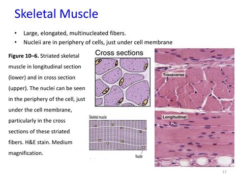
Table of Contents
Which Type of Muscle Tissue is Multinucleated?
Skeletal muscle tissue is the only type of muscle tissue that is multinucleated. This unique characteristic is crucial to its function and development, setting it apart from both smooth and cardiac muscle. Understanding the multinucleated nature of skeletal muscle requires exploring its development, structure, and the functional implications of this unique cellular architecture.
Understanding Multinucleated Cells
Before delving into the specifics of skeletal muscle, it's important to grasp the concept of multinucleation. Most cells in the human body are uninucleate, meaning they contain only one nucleus. The nucleus, as you likely know, houses the cell's genetic material (DNA), controlling all cellular activities. Multinucleated cells, however, contain multiple nuclei within a single cytoplasmic mass. This unusual cellular structure is not haphazard; it's a direct consequence of the cell's developmental pathway and its functional demands.
The presence of multiple nuclei allows for a significantly increased capacity for protein synthesis. This is particularly crucial in skeletal muscle cells, which require enormous quantities of proteins for contraction and repair. Each nucleus within a muscle fiber can independently transcribe genes and direct the production of proteins, greatly enhancing the overall efficiency of the cell. Imagine a single nucleus attempting to meet the protein demands of a large muscle fiber – it would simply be overwhelmed.
The Development of Multinucleated Skeletal Muscle Fibers
The multinucleated nature of skeletal muscle fibers is a direct result of their development. Unlike other cell types that arise from a single cell division, skeletal muscle fibers develop through a process called myogenesis. This intricate process involves several key steps:
1. Myoblast Formation and Proliferation:
Myogenesis begins with myoblasts, which are mononucleated precursor cells. These cells originate from the mesoderm, a germ layer during embryonic development. Myoblasts undergo rapid proliferation, increasing their numbers to meet the demand for muscle fiber formation.
2. Myoblast Fusion:
The defining event in myogenesis is the fusion of multiple myoblasts. This is a remarkable cellular process requiring precise molecular signaling and cell adhesion mechanisms. The membranes of multiple myoblasts fuse, resulting in a single, elongated cell containing multiple nuclei from the original myoblasts. This fusion process is not random; it's tightly regulated to ensure the proper arrangement and alignment of the resulting muscle fibers.
3. Myotube Formation and Maturation:
The fusion of myoblasts forms a myotube, an immature muscle fiber. The myotube undergoes a series of maturation processes, including the synthesis and assembly of contractile proteins (actin and myosin), the formation of myofibrils (the contractile units of muscle), and the development of the characteristic striated pattern of skeletal muscle. These processes are orchestrated by the coordinated actions of the multiple nuclei within the myotube, ensuring efficient protein production and organization.
4. Satellite Cells:
Even after the muscle fiber has matured, it retains a population of satellite cells. These are quiescent myoblasts located between the muscle fiber's plasma membrane and the basal lamina. Satellite cells play a crucial role in muscle growth, repair, and regeneration throughout an organism's lifespan. When a muscle is injured or undergoes hypertrophy (growth), satellite cells become activated, proliferate, and fuse with existing muscle fibers to repair damaged tissue or contribute to muscle growth. This process ensures that the multinucleated nature of skeletal muscle is maintained throughout life.
The Structure of Multinucleated Skeletal Muscle Fibers
A mature skeletal muscle fiber, also known as a muscle cell or myofiber, is a highly specialized, elongated, cylindrical cell that can be incredibly long (up to several centimeters). Its multinucleated structure is essential to its function. The nuclei are positioned just beneath the sarcolemma (plasma membrane) of the muscle fiber, which allows for efficient transport of mRNA and proteins to the myofibrils.
The muscle fiber's interior is highly organized, consisting of:
- Myofibrils: These are cylindrical structures running the length of the muscle fiber. They are the contractile units of the muscle, containing repeating units called sarcomeres.
- Sarcomeres: These are the basic functional units of muscle contraction. They are composed of precisely arranged actin and myosin filaments.
- Sarcoplasmic Reticulum (SR): This specialized endoplasmic reticulum is responsible for storing and releasing calcium ions (Ca2+), which are essential for muscle contraction.
- Transverse Tubules (T-tubules): These invaginations of the sarcolemma extend deep into the muscle fiber, allowing for rapid transmission of nerve impulses to the interior of the cell, triggering Ca2+ release from the SR and initiating muscle contraction.
The coordinated actions of these structural components, controlled by the multiple nuclei, allow for the powerful and precise contractions characteristic of skeletal muscle.
Functional Implications of Multinucleation in Skeletal Muscle
The multinucleated nature of skeletal muscle fibers offers several key advantages:
-
Increased Protein Synthesis Capacity: As mentioned earlier, the presence of multiple nuclei significantly increases the capacity for protein synthesis, which is crucial for muscle growth, repair, and the production of the contractile proteins required for efficient contraction.
-
Enhanced Metabolic Activity: The multiple nuclei also contribute to the enhanced metabolic activity of skeletal muscle fibers. Each nucleus can independently regulate gene expression, allowing for fine-tuning of metabolic pathways to meet the energy demands of the cell during both rest and activity.
-
Efficient Repair and Regeneration: The presence of satellite cells, which can fuse with existing muscle fibers, ensures efficient repair and regeneration of damaged muscle tissue. This is crucial for maintaining muscle function and preventing muscle degeneration.
-
Improved Coordination of Contraction: While the exact mechanism is still under investigation, the multiple nuclei might contribute to the coordinated contraction of the muscle fiber by allowing for the efficient distribution of signaling molecules and the precise regulation of contractile proteins within the sarcomeres.
Comparison with Other Muscle Tissue Types
In contrast to skeletal muscle, both smooth muscle and cardiac muscle are uninucleate. This difference reflects their distinct functions and developmental pathways.
-
Smooth Muscle: Found in the walls of internal organs and blood vessels, smooth muscle is responsible for involuntary movements such as peristalsis (the movement of food through the digestive tract) and vasoconstriction (the narrowing of blood vessels). Its uninucleated nature is sufficient for its slower, less forceful contractions.
-
Cardiac Muscle: Found in the heart, cardiac muscle is responsible for pumping blood throughout the body. Its uninucleated cells are interconnected through intercalated discs, which allow for rapid and coordinated contraction of the heart. While its contractions are more powerful than smooth muscle, they are still less powerful and less rapid than skeletal muscle contractions. The uninucleated nature of cardiac muscle is compatible with its rhythmic and synchronized contractions.
Conclusion: The Significance of Multinucleation in Skeletal Muscle
The multinucleated nature of skeletal muscle tissue is a unique and crucial aspect of its biology. This characteristic arises from the developmental process of myogenesis, where multiple myoblasts fuse to form a single, elongated muscle fiber containing multiple nuclei. This multinucleation significantly enhances the muscle fiber's capacity for protein synthesis, metabolic activity, repair, and overall function. In contrast to the uninucleated smooth and cardiac muscle tissues, the multinucleated skeletal muscle is perfectly adapted for its role in voluntary movement and its substantial power and speed of contraction. Understanding this fundamental difference is key to understanding the diverse functions of the various muscle tissue types within the human body. Further research continues to unravel the complexities of myogenesis and the precise roles of multiple nuclei in the sophisticated workings of skeletal muscle. The intricate interplay of genetics, cellular processes, and structural organization all contribute to the extraordinary power and adaptability of this remarkable tissue.
Latest Posts
Latest Posts
-
A Process By Which Information Is Exchanged Between Individuals
Apr 02, 2025
-
Select The Four Statements About Plasmodium That Are True
Apr 02, 2025
-
Greatest Common Factor Of 36 And 20
Apr 02, 2025
-
What Is The Antonym Of Urban
Apr 02, 2025
Related Post
Thank you for visiting our website which covers about Which Type Of Muscle Tissue Is Multinucleated . We hope the information provided has been useful to you. Feel free to contact us if you have any questions or need further assistance. See you next time and don't miss to bookmark.
