What Part Of The Ear Looks Like A Snail Shell
News Leon
Mar 17, 2025 · 6 min read
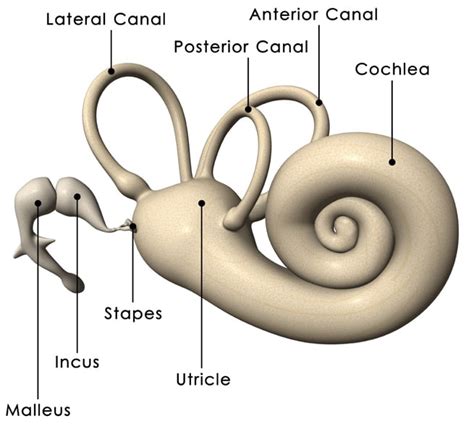
Table of Contents
What Part of the Ear Looks Like a Snail Shell? Exploring the Cochlea
The human ear, a marvel of biological engineering, is responsible for our sense of hearing and balance. While the external ear – the part we can see – is relatively straightforward in its appearance, the inner workings are far more complex and fascinating. One of the most intriguing structures within the inner ear is the cochlea, a remarkable spiral-shaped organ that truly resembles a snail shell. This article will delve deep into the anatomy, function, and significance of the cochlea, exploring its snail-like structure and its crucial role in our auditory experience.
The Cochlea: A Snail's Shell in Your Head
The cochlea, derived from the Greek word "κοχλίας" (cochlias) meaning "snail," is a fluid-filled, spiral-shaped cavity within the temporal bone of the skull. Its resemblance to a snail shell is not merely coincidental; this unique shape is critical to its function in processing sound. Imagine uncoiling this snail shell; you'd reveal a long, tapering tube approximately 3.5 centimeters in length. This tube is partitioned into three chambers:
The Three Chambers of the Cochlea
-
Scala Vestibuli: This is the uppermost chamber, connected to the oval window, a membrane-covered opening through which sound vibrations enter the inner ear. The oval window acts as a transducer, converting the mechanical vibrations from the middle ear into fluid waves within the cochlea.
-
Scala Media (Cochlear Duct): Located between the scala vestibuli and scala tympani, the scala media is the crucial chamber containing the Organ of Corti, the sensory organ of hearing. It's filled with endolymph, a fluid with a unique ionic composition different from the perilymph found in the other two chambers. This difference in ionic composition is essential for the generation of electrical signals.
-
Scala Tympani: This is the lowermost chamber, connected to the round window, a membrane-covered opening that allows the fluid in the cochlea to move in response to the sound vibrations. The round window acts as a pressure release valve, preventing excessive pressure buildup within the cochlea. This pressure regulation is essential for efficient sound transmission.
The Organ of Corti: The Heart of Hearing
Nestled within the scala media lies the Organ of Corti, a remarkable structure responsible for converting the fluid vibrations into electrical signals that the brain interprets as sound. This organ is incredibly intricate, comprising a complex array of hair cells, supporting cells, and the basilar membrane.
Hair Cells: The Sensory Receptors of Hearing
The hair cells are the sensory receptors of hearing, responsible for transducing the mechanical vibrations into electrical signals. There are two main types of hair cells:
-
Inner Hair Cells (IHCs): These are arranged in a single row and are primarily responsible for transmitting auditory information to the brain. They are the main contributors to our perception of sound.
-
Outer Hair Cells (OHCs): Arranged in three rows, these cells play a crucial role in amplifying faint sounds and enhancing the sharpness of our hearing. They act as tiny adjustable amplifiers, sharpening the frequency response of the cochlea.
Basilar Membrane: The Frequency Analyzer
The basilar membrane is a flexible membrane running the length of the cochlea. It's wider and more flexible at the apex (the tip of the snail shell) and narrower and stiffer at the base (near the oval window). This variation in stiffness is critical for frequency discrimination. Different frequencies stimulate different regions of the basilar membrane:
-
High-frequency sounds: These stimulate the basilar membrane near the base, where it's stiffer.
-
Low-frequency sounds: These stimulate the basilar membrane near the apex, where it's more flexible.
This tonotopic organization, where different frequencies are mapped to different locations along the basilar membrane, is essential for our ability to distinguish between various sounds. The hair cells located at the point of maximal displacement of the basilar membrane send the strongest signals to the brain, allowing us to perceive the frequency of the sound.
The Journey of Sound: From Ear to Brain
The process of hearing begins when sound waves enter the outer ear and travel down the auditory canal to the eardrum (tympanic membrane). The vibrations of the eardrum are then amplified by the three tiny bones in the middle ear (malleus, incus, and stapes). The stapes transmits these vibrations to the oval window, initiating the process within the inner ear.
The fluid waves generated in the cochlea stimulate the hair cells on the basilar membrane, causing them to release neurotransmitters. This triggers electrical signals in the auditory nerve fibers, which transmit the information to the brainstem. The brainstem then relays these signals to the auditory cortex in the temporal lobe of the brain, where the sound is finally interpreted.
The Cochlea and Hearing Loss
Damage to the cochlea, whether due to aging, noise exposure, disease, or genetics, can lead to various types of hearing loss. Since the cochlea is responsible for converting sound vibrations into electrical signals, damage to this structure directly impacts the ability to hear. Different types of hearing loss can affect various parts of the cochlea and result in different hearing impairments:
-
Sensorineural hearing loss: This is the most common type of hearing loss and results from damage to the hair cells or auditory nerve. This damage can be caused by prolonged exposure to loud noise, aging, certain medications, and genetic disorders.
-
Conductive hearing loss: This type of hearing loss occurs when sound waves are unable to reach the inner ear effectively. This is often due to problems in the outer or middle ear, such as earwax buildup, middle ear infections, or damage to the ossicles.
-
Mixed hearing loss: This is a combination of sensorineural and conductive hearing loss.
Cochlear Implants: Restoring Hearing
For individuals with severe sensorineural hearing loss, cochlear implants offer a remarkable technological solution. These devices bypass the damaged hair cells and directly stimulate the auditory nerve. A cochlear implant consists of an external speech processor and an internal implant surgically placed within the cochlea. The speech processor converts sounds into electrical signals, which are then transmitted to the electrodes within the cochlea, stimulating the auditory nerve and allowing the brain to perceive sound.
Conclusion: The Snail Shell That Enables Hearing
The cochlea, with its remarkable snail-shell structure, is a critical component of the human auditory system. Its intricate anatomy and complex mechanisms allow us to perceive a wide range of sounds, from the softest whispers to the loudest thunder. Understanding the cochlea's structure and function is crucial not only for appreciating the wonder of our hearing but also for developing effective treatments for hearing loss. Further research continues to unravel the complexities of this fascinating organ and its vital role in our perception of the world. The ongoing exploration of the cochlea highlights the intricate and elegant design of the human body and the constant pursuit of scientific advancement in the field of audiology. From its spiral shape to its intricate inner workings, the cochlea stands as a testament to the remarkable capabilities of biological engineering, constantly inspiring wonder and further research.
Latest Posts
Latest Posts
-
Which Of The Following Substances Has The Highest Melting Point
Mar 17, 2025
-
Mountain Range Separating Europe From Asia
Mar 17, 2025
-
200 Cm Equals How Many M
Mar 17, 2025
-
How Many Yards In 90 Inches
Mar 17, 2025
-
Explain Why Plants Are Called Producers
Mar 17, 2025
Related Post
Thank you for visiting our website which covers about What Part Of The Ear Looks Like A Snail Shell . We hope the information provided has been useful to you. Feel free to contact us if you have any questions or need further assistance. See you next time and don't miss to bookmark.
