What Is The Pattern Of Microtubule Arrangement In A Centriole
News Leon
Mar 24, 2025 · 6 min read
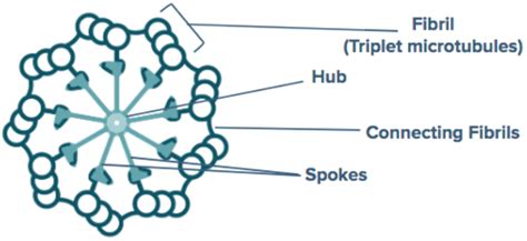
Table of Contents
What is the Pattern of Microtubule Arrangement in a Centriole?
Centrioles are essential, cylindrical organelles found in most eukaryotic cells. They play a crucial role in cell division, organization of the cytoskeleton, and various other cellular processes. Understanding their structure, particularly the precise arrangement of microtubules within them, is fundamental to comprehending their function. This article will delve into the intricate pattern of microtubule arrangement in a centriole, exploring its significance and the implications of any deviations from the standard structure.
The Nine-Triplet Microtubule Structure: A Defining Characteristic
The defining characteristic of a centriole is its highly organized array of microtubules. Unlike other microtubule-based structures, centrioles feature a 9+0 arrangement. This means nine triplet microtubules are arranged in a ring, surrounding a hollow center devoid of any microtubules (hence the '0'). This distinctive pattern is strikingly conserved across a wide range of eukaryotic species, highlighting its fundamental importance.
Understanding Microtubule Triplets
Each of these nine triplet microtubules is composed of three individual microtubules – A, B, and C. Microtubule A is a complete microtubule, meaning it possesses a full complement of 13 protofilaments. Microtubules B and C, however, are incomplete, sharing protofilaments with microtubule A. Microtubule B shares approximately three protofilaments with A, while microtubule C shares approximately three protofilaments with B. This intricate sharing of protofilaments contributes to the structural integrity and stability of the centriole.
The Role of Microtubule-Associated Proteins (MAPs)
The precise arrangement of these microtubules isn't merely a consequence of self-assembly. Numerous microtubule-associated proteins (MAPs) play a vital role in the construction and maintenance of the centriolar structure. These proteins act as scaffolds, ensuring the accurate alignment and stable interaction between the microtubules. Specific MAPs are crucial for:
- Nucleation: Initiating the polymerization of tubulin dimers into microtubules.
- Anchoring: Securely linking the microtubules to each other and to the centriolar matrix.
- Organization: Guiding the precise arrangement of the microtubule triplets.
- Regulation: Controlling the dynamics of microtubule assembly and disassembly.
The precise identity and function of many of these MAPs are still under investigation, but their importance in the centriole’s structural integrity and functionality is undeniable.
Variations and Deviations from the Canonical 9+0 Arrangement
While the 9+0 arrangement is considered the canonical structure, variations do exist. These variations can occur due to:
- Species-Specific Differences: Some organisms exhibit subtle differences in the precise arrangement of microtubules or the associated proteins. These variations may reflect evolutionary adaptations or functional specializations.
- Developmental Stages: The organization of microtubules within centrioles can change during different developmental stages of an organism or even within a single cell cycle.
- Pathological Conditions: Disruptions in the normal microtubule arrangement can be indicative of various cellular pathologies, including cancer and developmental disorders.
These deviations, though sometimes subtle, can profoundly impact the functionality of the centriole and the cell as a whole.
Consequences of Aberrant Microtubule Arrangement
Any deviation from the precise 9+0 structure can have significant consequences. These can include:
- Defects in Cell Division: Accurate chromosome segregation during mitosis and meiosis relies heavily on the precise organization of the mitotic spindle, which is nucleated by the centrioles. Aberrant centriolar structure can lead to chromosome mis-segregation, resulting in aneuploidy and potential cell death.
- Impaired Cilia and Flagella Formation: Centrioles serve as basal bodies for cilia and flagella, essential structures involved in cell motility and sensory functions. Defective centrioles can result in dysfunctional cilia and flagella, leading to various genetic disorders.
- Cytoskeletal Instability: Centrioles contribute to the overall organization of the cytoskeleton, influencing cell shape, intracellular transport, and cell polarity. Disruptions in centriolar structure can lead to cytoskeletal instability, potentially affecting a range of cellular processes.
- Cancer Development: Aberrations in centriole structure and number (often resulting in increased centriole numbers, known as centriole amplification) have been implicated in the development and progression of various cancers.
Methods for Studying Centriole Microtubule Arrangement
Investigating the intricate details of centriole microtubule arrangement requires advanced microscopic and biochemical techniques. Some of the most commonly used methods include:
- Electron Microscopy (EM): EM, particularly transmission electron microscopy (TEM), provides high-resolution images allowing visualization of individual microtubules and their interactions. This is crucial for confirming the 9+0 arrangement and identifying any deviations.
- Immunofluorescence Microscopy: Using specific antibodies against tubulin and other centriolar proteins, researchers can visualize the microtubules and associated proteins within the centriole, providing insight into their arrangement and interactions.
- Cryo-Electron Tomography (Cryo-ET): This technique allows for three-dimensional reconstruction of the centriole at near-atomic resolution, providing a detailed understanding of the structural organization and the interactions between microtubules and associated proteins.
- Biochemical Assays: Techniques such as co-immunoprecipitation and protein interaction mapping can be used to identify the proteins associated with the centriole and determine their interactions.
These techniques, often used in combination, allow for a comprehensive understanding of the centriole's structure and function.
The Significance of the 9+0 Pattern: A Conservation Mystery
The remarkable conservation of the 9+0 pattern across diverse eukaryotic species suggests a fundamental importance of this structure. The precise reasons for this highly conserved arrangement remain a subject of ongoing research, but several hypotheses exist:
- Mechanical Strength and Stability: The 9+0 arrangement provides exceptional structural strength and stability to the centriole, allowing it to withstand the forces encountered during cell division and other cellular processes.
- Precise Organization of the Mitotic Spindle: The specific arrangement of microtubules is crucial for accurately organizing the mitotic spindle, ensuring precise chromosome segregation.
- Efficient Nucleation of Microtubules: The centriole's structure may facilitate efficient nucleation of microtubules, promoting the rapid assembly of the mitotic spindle.
- Regulation of Microtubule Dynamics: The centriole may play a role in regulating the dynamics of microtubules, controlling their assembly and disassembly, and ensuring the proper organization of the cytoskeleton.
Further research is needed to fully elucidate the functional significance of this unique and highly conserved structure.
Conclusion: Unveiling the Secrets of the Centriole
The precise pattern of microtubule arrangement in a centriole – the defining 9+0 structure – is far more than just an anatomical feature. It represents a sophisticated and highly conserved architectural marvel, crucial for a wide range of cellular functions. Understanding this arrangement, the associated proteins, and the consequences of any deviations is paramount for comprehending fundamental biological processes and tackling diseases associated with centriolar dysfunction. Continued research, employing advanced imaging and biochemical techniques, will further unravel the secrets held within this remarkably conserved organelle and its crucial role in cell biology. Further investigations into the specific MAPs involved, the dynamic nature of the centriole throughout the cell cycle, and the precise mechanisms underpinning its remarkable structural stability are essential to fully appreciate the significance of this amazing cellular component. The quest to completely understand the centriole and its microtubule arrangement remains an active and exciting area of ongoing research, with implications extending far beyond basic cell biology into the realms of medicine and biotechnology.
Latest Posts
Latest Posts
-
A Pectoral Girdle Consists Of Two Bones The And The
Mar 28, 2025
-
Separation Of Homologous Chromosomes Occurs During
Mar 28, 2025
-
There Are Pairs Of True Ribs
Mar 28, 2025
-
Salivary Amylase Begins The Digestion Of
Mar 28, 2025
-
When A Cell Is Placed In A Hypertonic Solution
Mar 28, 2025
Related Post
Thank you for visiting our website which covers about What Is The Pattern Of Microtubule Arrangement In A Centriole . We hope the information provided has been useful to you. Feel free to contact us if you have any questions or need further assistance. See you next time and don't miss to bookmark.
