What Is The Functional Unit Of A Skeletal Muscle Called
News Leon
Mar 17, 2025 · 6 min read
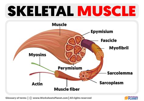
Table of Contents
What is the Functional Unit of a Skeletal Muscle Called?
The question, "What is the functional unit of a skeletal muscle called?" has a straightforward answer: the sarcomere. However, understanding the sarcomere's function requires delving into the intricate structure and mechanics of skeletal muscle. This article will explore the sarcomere in detail, explaining its composition, how it contracts, and its significance in overall muscle function. We'll also touch upon related concepts like muscle fibers, myofibrils, and the neuromuscular junction to provide a complete picture of skeletal muscle physiology.
Understanding Skeletal Muscle Structure
Before diving into the sarcomere, let's establish a foundational understanding of skeletal muscle's hierarchical organization. Skeletal muscle, responsible for voluntary movement, is composed of numerous structural components, each nested within the next:
1. Muscle Fibers: The Building Blocks
Skeletal muscles are made up of elongated, cylindrical cells called muscle fibers or myofibers. These fibers are multinucleated, meaning they contain multiple nuclei within their cytoplasm (sarcoplasm). This multinucleated nature reflects their development from the fusion of multiple myoblasts during embryogenesis. The sarcolemma, the muscle fiber's plasma membrane, plays a crucial role in transmitting signals for muscle contraction.
2. Myofibrils: The Contractile Machines
Within each muscle fiber, numerous myofibrils run parallel to the long axis. These cylindrical structures are the actual contractile units of the muscle fiber. They are densely packed with highly organized protein filaments, primarily actin and myosin, responsible for the muscle's ability to generate force.
3. Sarcomeres: The Functional Units
Finally, we arrive at the sarcomere, the fundamental unit of contraction within the myofibril. This highly organized structure, repeating along the length of the myofibril, is responsible for the characteristic striated appearance of skeletal muscle under a microscope. The sarcomere's structure and function are intimately linked, with precise arrangement of its components crucial for efficient muscle contraction.
The Sarcomere: A Detailed Look
The sarcomere is defined by two Z-lines (or Z-discs), which are dense protein structures that serve as attachment points for the thin filaments (primarily actin). The region between two consecutive Z-lines constitutes a single sarcomere.
Key Components of the Sarcomere:
-
Z-lines: As mentioned, these define the boundaries of the sarcomere. They anchor the thin filaments.
-
Thin Filaments (Actin): Composed primarily of the protein actin, these filaments extend from the Z-lines toward the center of the sarcomere. They also contain two other important proteins: tropomyosin, which wraps around the actin filament, and troponin, which sits on tropomyosin and plays a crucial role in regulating muscle contraction.
-
Thick Filaments (Myosin): These filaments are located in the center of the sarcomere and are composed of the protein myosin. Each myosin molecule has a head and a tail, and the heads are responsible for binding to actin and generating force during contraction.
-
A-band: This is the dark band seen under a microscope, representing the region where both thick and thin filaments overlap.
-
I-band: This is the light band, containing only thin filaments. It bisects the Z-line.
-
H-zone: This is a lighter region within the A-band, containing only thick filaments.
-
M-line: This is a protein structure located in the center of the H-zone, anchoring the thick filaments.
The Sliding Filament Theory: How Sarcomeres Contract
Muscle contraction occurs through the sliding filament theory. This theory explains how the thin and thick filaments slide past each other, shortening the sarcomere and ultimately the entire muscle fiber.
The process is initiated by a nerve impulse at the neuromuscular junction, the specialized synapse between a motor neuron and a muscle fiber. This impulse triggers the release of acetylcholine, a neurotransmitter that depolarizes the sarcolemma and initiates a cascade of events within the muscle fiber.
The depolarization spreads through the T-tubules, invaginations of the sarcolemma that extend deep into the muscle fiber, triggering the release of calcium ions (Ca2+) from the sarcoplasmic reticulum, a specialized intracellular calcium store.
The released calcium ions bind to troponin on the thin filaments, causing a conformational change in tropomyosin that exposes the myosin-binding sites on actin. This allows the myosin heads to bind to actin, forming cross-bridges.
The myosin heads then undergo a power stroke, pulling the thin filaments toward the center of the sarcomere. This process requires ATP (adenosine triphosphate), the energy currency of the cell. The myosin heads detach from actin, re-cock, and reattach, repeating the cycle. This continuous cycle of cross-bridge formation, power strokes, and detachment leads to the shortening of the sarcomere.
As the thin filaments slide past the thick filaments, the I-band and H-zone narrow, while the A-band remains relatively constant in length. The overall effect is a shortening of the sarcomere and, consequently, the muscle fiber and the entire muscle.
When the nerve impulse ceases, calcium ions are actively pumped back into the sarcoplasmic reticulum, causing tropomyosin to block the myosin-binding sites on actin. Muscle contraction stops, and the muscle fiber relaxes.
Sarcomere Function in Different Muscle Fiber Types
While the basic sarcomere structure and contraction mechanism are similar across all skeletal muscle fibers, there are variations in the expression of different protein isoforms and the fiber's metabolic properties, leading to different muscle fiber types.
Type I (Slow-twitch) fibers: These fibers are specialized for endurance activities, characterized by a slow rate of contraction and high resistance to fatigue. They have a high density of mitochondria, providing a sustainable energy supply via oxidative phosphorylation.
Type IIa (Fast-twitch oxidative) fibers: These fibers exhibit a faster contraction rate than Type I fibers and are moderately resistant to fatigue. They use a combination of oxidative and glycolytic metabolism.
Type IIb (Fast-twitch glycolytic) fibers: These fibers contract rapidly but fatigue quickly. They primarily rely on anaerobic glycolysis for energy production.
The differences in the contractile properties of these fiber types are partly determined by the differences in the isoform of myosin present, the density of mitochondria and capillaries, and the efficiency of calcium handling by the sarcoplasmic reticulum.
Clinical Significance of Sarcomere Dysfunction
Disruptions to sarcomere function can lead to a variety of muscle disorders. These can be genetic or acquired.
Examples of sarcomere-related diseases include:
-
Muscular dystrophies: These genetic disorders affect the structure and function of muscle proteins, leading to progressive muscle weakness and degeneration. Examples include Duchenne muscular dystrophy and Becker muscular dystrophy.
-
Inherited cardiomyopathies: These are heart muscle diseases that can involve mutations in genes encoding sarcomeric proteins. This can lead to heart failure and other cardiac complications.
-
Rhabdomyolysis: This condition is characterized by the breakdown of skeletal muscle fibers, releasing muscle proteins into the bloodstream. It can be triggered by strenuous exercise, trauma, or certain medications.
Understanding the sarcomere's structure and function is crucial for comprehending normal muscle physiology and the pathogenesis of various muscle disorders. Further research continues to unveil the intricate mechanisms of sarcomere function and the development of new therapeutic strategies for muscle-related diseases.
Conclusion
In summary, the functional unit of a skeletal muscle is the sarcomere. This highly organized structure, comprised of actin and myosin filaments, enables muscle contraction via the sliding filament theory. The sarcomere's intricate arrangement and the precise interplay of its components are essential for generating force and movement. Variations in sarcomere composition and function contribute to the diversity of muscle fiber types, determining their contractile properties and resistance to fatigue. Furthermore, disruptions in sarcomere function underlie a wide range of muscle disorders, highlighting the clinical importance of understanding this fundamental unit of muscle physiology. Continued research on sarcomeres promises to improve our understanding of muscle health and disease.
Latest Posts
Latest Posts
-
Which Of The Following Is Not A Form Of Precipitation
Mar 18, 2025
-
Which Statement About Natural Selection Is True
Mar 18, 2025
-
Which Chamber Of Heart Has Thickest Wall
Mar 18, 2025
-
How Many Feet Is 1 2 Miles
Mar 18, 2025
-
How Many Valence Electrons Does Mn Have
Mar 18, 2025
Related Post
Thank you for visiting our website which covers about What Is The Functional Unit Of A Skeletal Muscle Called . We hope the information provided has been useful to you. Feel free to contact us if you have any questions or need further assistance. See you next time and don't miss to bookmark.
