What Is The Functional Contractile Unit Of The Myofibril
News Leon
Mar 15, 2025 · 7 min read
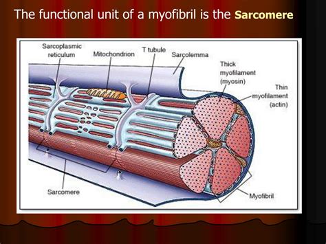
Table of Contents
What is the Functional Contractile Unit of the Myofibril?
The human body is a marvel of engineering, capable of a breathtaking array of movements, from the delicate tap of a finger to the powerful sprint of a marathon runner. This intricate capacity for movement is fundamentally driven by the sophisticated machinery housed within our muscles: the sarcomeres. Understanding the sarcomere, the functional contractile unit of the myofibril, is key to understanding how muscles work and what factors influence their performance.
Delving into the Myofibril: A Microscopic Look at Muscle Contraction
Before diving into the specifics of the sarcomere, it's crucial to establish its context within the larger muscular structure. Muscles are composed of bundles of muscle fibers, which in turn are comprised of numerous elongated cylindrical structures called myofibrils. These myofibrils are the fundamental units of muscle contraction, responsible for the actual shortening and lengthening of the muscle. Within the myofibril, a highly organized and repetitive pattern of protein filaments creates the foundation for the muscle's contractile power. This pattern is visible under a microscope as alternating light and dark bands, a characteristic striation that gives skeletal and cardiac muscle their name. These bands reflect the precise arrangement of the protein filaments that comprise the sarcomere.
The Significance of Striations: A Visual Cue to Contractile Function
The striated appearance of skeletal and cardiac muscle fibers results directly from the precise arrangement of two key proteins: actin and myosin. These filaments are not randomly distributed; they are organized into highly structured units that repeat along the length of the myofibril. These repeating units are the sarcomeres, the fundamental units responsible for muscle contraction.
The Sarcomere: Anatomy and Function of the Contractile Unit
The sarcomere is defined by specific boundaries formed by protein structures. Let's examine its key components:
1. Z-lines (Z-discs): The Defining Boundaries
The Z-lines (or Z-discs) are dense protein structures that mark the boundaries of each sarcomere. They are crucial for anchoring the thin filaments (actin) and play a vital role in the transmission of force during contraction. The distance between two consecutive Z-lines defines the length of the sarcomere.
2. I-bands: Light Bands Representing Actin Filaments
The I-bands (isotropic bands) are the lighter areas of the sarcomere. These bands contain only thin filaments (primarily actin), extending from the Z-line towards the center of the sarcomere, but not overlapping with the thick filaments. The I-bands shorten during muscle contraction as the thin filaments slide inward.
3. A-bands: Dark Bands Representing Myosin Filaments
The A-bands (anisotropic bands) are the darker areas of the sarcomere. These bands contain the thick filaments (primarily myosin), which are significantly longer than the thin filaments. The A-bands remain relatively constant in length during muscle contraction, even though the overall sarcomere shortens.
4. H-zone: The Myosin-Only Region
Within the A-band, there's a lighter central region called the H-zone (Hensen's zone). This zone contains only the thick filaments (myosin), devoid of overlapping thin filaments. The H-zone narrows during muscle contraction as the thin filaments slide inward, overlapping increasingly with the thick filaments.
5. M-line: The Center of the Sarcomere
The M-line (middle line) is located in the center of the H-zone and serves as an anchoring point for the myosin filaments. It helps to maintain the structural integrity of the sarcomere and plays a role in organizing the myosin molecules within the thick filaments.
The Sliding Filament Theory: How Sarcomeres Contract
The process of muscle contraction is elegantly explained by the sliding filament theory. This theory posits that muscle contraction occurs due to the sliding of the thin (actin) filaments past the thick (myosin) filaments, thereby shortening the sarcomere without altering the length of the individual filaments themselves.
The Role of Myosin Heads: The Engines of Contraction
The thick filaments (myosin) have globular heads that project outwards and interact with the thin filaments (actin). These myosin heads possess ATPase activity, meaning they can break down adenosine triphosphate (ATP) to release energy. This energy fuels the cyclical interaction between the myosin heads and the actin filaments.
The Cross-Bridge Cycle: A Repeating Sequence of Interactions
The myosin heads bind to specific sites on the actin filaments, forming cross-bridges. This binding is facilitated by calcium ions (Ca²⁺), which are released into the sarcoplasm (the cytoplasm of muscle fibers) upon stimulation from a nerve impulse. Once bound, the myosin heads undergo a conformational change, pulling the thin filaments towards the center of the sarcomere. This "power stroke" requires the energy released from ATP hydrolysis. After the power stroke, the myosin head detaches from the actin filament, resets to its original conformation, and then binds to a new site further along the actin filament. This cycle repeats numerous times, leading to the progressive sliding of the thin filaments and shortening of the sarcomere.
The Significance of Calcium Ions: Regulating Muscle Contraction
Calcium ions (Ca²⁺) are essential for muscle contraction. Their presence allows for the interaction between the myosin heads and the actin filaments. In the absence of Ca²⁺, a protein called tropomyosin blocks the myosin-binding sites on the actin filaments, preventing the cross-bridge cycle from occurring. When a nerve impulse stimulates the muscle fiber, calcium ions are released, binding to troponin, a protein complex associated with tropomyosin. This binding causes a conformational change in tropomyosin, exposing the myosin-binding sites on the actin filaments, thus initiating the cross-bridge cycle and muscle contraction.
Factors Affecting Sarcomere Function and Muscle Performance
Several factors influence the function of the sarcomere and ultimately the performance of the muscle:
1. Sarcomere Length: Optimal Overlap for Maximum Force
The length of the sarcomere at the onset of contraction significantly impacts the force it can generate. An optimal sarcomere length allows for maximal overlap between the actin and myosin filaments, resulting in maximum cross-bridge formation and force production. Both excessively short and excessively long sarcomeres reduce the number of potential cross-bridges and therefore decrease the force generated.
2. ATP Availability: Fueling Muscle Contraction
ATP is crucial for the cross-bridge cycle and muscle contraction. A depletion of ATP leads to muscle fatigue and inability to continue contracting. The body employs different metabolic pathways to generate ATP, including aerobic respiration and anaerobic glycolysis, depending on the intensity and duration of muscle activity.
3. Calcium Ion Concentration: Fine-Tuning Contraction
The concentration of calcium ions (Ca²⁺) in the sarcoplasm is meticulously controlled. Precise regulation of Ca²⁺ levels is critical for both initiating and terminating muscle contraction. Sustained elevated Ca²⁺ levels can lead to muscle cramps and fatigue.
4. Muscle Fiber Type: Different Contractile Properties
Muscles contain different types of muscle fibers, each exhibiting unique contractile properties. Type I (slow-twitch) fibers are specialized for endurance activities, while Type II (fast-twitch) fibers are adapted for rapid, powerful contractions. These differences in fiber type reflect variations in sarcomere structure and metabolic capabilities.
5. Age and Disease: Factors Affecting Sarcomere Integrity
The age and overall health of an individual influence the integrity and function of the sarcomere. Age-related changes, such as sarcopenia (loss of muscle mass), and diseases like muscular dystrophy can impair sarcomere function, leading to reduced muscle strength and impaired mobility.
Conclusion: The Sarcomere – A Masterpiece of Biological Engineering
The sarcomere, the functional contractile unit of the myofibril, is a masterpiece of biological engineering. Its highly organized structure and precise molecular interactions enable the remarkable ability of muscles to generate force and movement. Understanding the intricacies of the sarcomere and its role in muscle contraction is fundamental to comprehending human movement, athletic performance, and the impact of age and disease on musculoskeletal health. Further research into sarcomere function promises to unlock innovative therapeutic approaches for treating muscle disorders and enhancing physical capabilities. From the microscopic world of proteins to the macroscopic realm of human movement, the sarcomere remains a captivating subject of study, continuing to reveal its secrets to those who delve into the depths of its fascinating mechanics.
Latest Posts
Latest Posts
-
In The Figure The Electric Field Lines On The Left
Mar 15, 2025
-
What Is The Least Common Multiple Of 4 And 9
Mar 15, 2025
-
Iodine Is Essential For The Synthesis Of
Mar 15, 2025
-
Is Osmosis High To Low Or Low To High
Mar 15, 2025
-
Concave Mirror And Convex Mirror Difference
Mar 15, 2025
Related Post
Thank you for visiting our website which covers about What Is The Functional Contractile Unit Of The Myofibril . We hope the information provided has been useful to you. Feel free to contact us if you have any questions or need further assistance. See you next time and don't miss to bookmark.
