The Small Space Between Neurons Is Called ____________________.
News Leon
Mar 21, 2025 · 6 min read
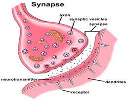
Table of Contents
The Small Space Between Neurons is Called a Synapse: A Deep Dive into Neural Communication
The small space between neurons is called a synapse. This seemingly minuscule gap plays a pivotal role in the intricate workings of the nervous system, acting as the crucial site of communication between nerve cells. Understanding the synapse is key to unlocking the mysteries of the brain, from simple reflexes to complex cognitive functions. This article will explore the structure, function, and significance of the synapse, delving into the fascinating world of neuronal communication.
Understanding the Structure of a Synapse
A synapse is far more than just an empty space; it's a highly specialized junction meticulously designed to facilitate the transmission of signals. It's comprised of three main components:
1. The Presynaptic Neuron: The Message Sender
This is the neuron sending the signal. At the end of its axon, the presynaptic neuron possesses specialized structures called synaptic boutons or terminal buttons. These boutons are packed with synaptic vesicles, tiny sacs containing neurotransmitters, the chemical messengers of the nervous system. These neurotransmitters are the key players in transmitting information across the synaptic cleft. The presynaptic neuron's membrane also contains voltage-gated calcium channels, crucial for the release of neurotransmitters.
2. The Synaptic Cleft: The Communication Bridge
The synaptic cleft is the actual space between the presynaptic and postsynaptic neurons. This gap, typically measuring only 20-40 nanometers (billionths of a meter) wide, is not simply empty. It's a precisely regulated microenvironment containing extracellular fluid and various molecules that influence neurotransmitter activity. The small size of the cleft ensures efficient neurotransmitter diffusion and rapid signal transmission.
3. The Postsynaptic Neuron: The Message Receiver
This is the neuron receiving the signal. Its membrane directly opposite the presynaptic terminal contains specialized receptor proteins. These receptors are uniquely shaped to bind with specific neurotransmitters. Upon binding, these receptors trigger a cascade of events within the postsynaptic neuron, either exciting or inhibiting its activity. The postsynaptic membrane also contains various other proteins involved in signal transduction and synaptic plasticity.
The Process of Synaptic Transmission: A Chemical Dance
The transmission of signals across the synapse is a complex, multi-step process:
1. Action Potential Arrival: The Trigger
An electrical signal, known as an action potential, travels down the axon of the presynaptic neuron. This electrical impulse reaches the axon terminal, initiating the release of neurotransmitters.
2. Calcium Influx: The Gate Opener
The arrival of the action potential triggers the opening of voltage-gated calcium channels in the presynaptic membrane. Calcium ions (Ca²⁺) rush into the axon terminal, a critical step in triggering neurotransmitter release.
3. Vesicle Fusion and Neurotransmitter Release: The Messenger's Departure
The influx of calcium ions causes the synaptic vesicles to fuse with the presynaptic membrane, releasing their neurotransmitter contents into the synaptic cleft through a process called exocytosis.
4. Neurotransmitter Diffusion and Receptor Binding: The Message Across the Gap
The released neurotransmitters diffuse across the synaptic cleft and bind to their specific receptors on the postsynaptic membrane. This binding initiates a change in the postsynaptic neuron's membrane potential.
5. Postsynaptic Potential: Excitation or Inhibition
The binding of neurotransmitters can cause either an excitatory postsynaptic potential (EPSP) or an inhibitory postsynaptic potential (IPSP). EPSPs depolarize the postsynaptic membrane, bringing it closer to the threshold for firing an action potential, while IPSPs hyperpolarize the membrane, making it less likely to fire.
6. Neurotransmitter Removal: Resetting the System
After the neurotransmitter has exerted its effect, it needs to be removed from the synaptic cleft to prevent continuous stimulation. This removal occurs through several mechanisms: reuptake by the presynaptic neuron, enzymatic degradation, and diffusion away from the synapse.
Types of Synapses: Variations on a Theme
Synapses are not all created equal. They can be categorized based on several criteria:
1. Based on the Type of Signal Transmitted:
-
Chemical Synapses: These are the most common type of synapse, relying on the release of neurotransmitters to transmit signals across the cleft. The process described above is characteristic of chemical synapses.
-
Electrical Synapses: In these synapses, the pre- and postsynaptic neurons are directly connected by gap junctions, allowing for the direct flow of electrical current. Electrical synapses are faster than chemical synapses but offer less flexibility in signal modulation.
2. Based on the Location of the Synapse:
-
Axodendritic Synapses: The synapse occurs between the axon of the presynaptic neuron and the dendrite of the postsynaptic neuron. This is the most common type.
-
Axosomatic Synapses: The synapse occurs between the axon of the presynaptic neuron and the soma (cell body) of the postsynaptic neuron.
-
Axoaxonic Synapses: The synapse occurs between the axon of one neuron and the axon of another. These synapses often modulate the release of neurotransmitters from the postsynaptic axon.
Synaptic Plasticity: The Adaptable Synapse
Synapses are not static structures; their strength and efficacy can change over time, a phenomenon known as synaptic plasticity. This plasticity is crucial for learning and memory. Long-term potentiation (LTP) and long-term depression (LTD) are two prominent forms of synaptic plasticity, involving long-lasting changes in synaptic strength.
The Significance of Synapses in Neurological Disorders
Dysfunction in synaptic transmission is implicated in numerous neurological and psychiatric disorders. For example:
-
Alzheimer's Disease: The formation of amyloid plaques and neurofibrillary tangles disrupts synaptic function, leading to cognitive decline.
-
Parkinson's Disease: Degeneration of dopaminergic neurons in the substantia nigra affects synaptic transmission in the basal ganglia, resulting in motor impairments.
-
Schizophrenia: Imbalances in neurotransmitter systems, particularly dopamine and glutamate, are believed to contribute to the symptoms of schizophrenia.
-
Depression: Dysregulation of neurotransmitters like serotonin and norepinephrine is implicated in the pathogenesis of depression.
Research and Future Directions
Research into synaptic function and plasticity is ongoing, with significant advancements constantly being made. Techniques like optogenetics and advanced imaging methods are providing unprecedented insights into the intricate mechanisms of synaptic transmission. Understanding the intricacies of the synapse holds the key to developing more effective treatments for neurological and psychiatric disorders. Further research will likely focus on:
-
Developing novel drugs that target specific aspects of synaptic transmission. This may involve modulating neurotransmitter release, receptor activity, or the processes involved in synaptic plasticity.
-
Exploring the role of glial cells in synaptic function. Glial cells, once considered mere support cells, are now recognized as active players in synaptic transmission and plasticity.
-
Investigating the complex interactions between different neurotransmitter systems. Neurotransmitter systems are not isolated entities; they interact in complex ways to shape neuronal activity.
-
Developing more sophisticated computational models of synaptic function. These models can help us understand the emergent properties of neural networks and predict the effects of various interventions.
Conclusion: The Synapse - A Tiny Space, A Mighty Influence
The small space between neurons, the synapse, is far from insignificant. It is the site of dynamic communication, the foundation of neural computation, and a key player in our cognitive abilities. From the precise molecular interactions to the complex interplay of neurotransmitters and receptors, the synapse remains a fascinating and essential area of neuroscience research. Continued investigation into its intricacies promises to unravel further mysteries of the brain and lead to advancements in treating a wide range of neurological and psychiatric conditions. The seemingly simple gap between neurons is, in reality, a microcosm of the complexity and wonder of the nervous system.
Latest Posts
Latest Posts
-
Do Noble Gases Have Ionization Energy
Mar 27, 2025
-
How To Write An Invitation Letter To A Friend
Mar 27, 2025
-
The System In The Figure Below Is In Equilibrium
Mar 27, 2025
-
Poisonous Substances Produced By Some Microorganisms Are Called
Mar 27, 2025
-
In What Cell Organelle Does Photosynthesis Occur
Mar 27, 2025
Related Post
Thank you for visiting our website which covers about The Small Space Between Neurons Is Called ____________________. . We hope the information provided has been useful to you. Feel free to contact us if you have any questions or need further assistance. See you next time and don't miss to bookmark.
