The Joints Between Cranial Bones Of The Skull Are Called...
News Leon
Mar 19, 2025 · 6 min read
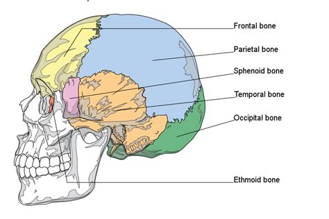
Table of Contents
The Joints Between Cranial Bones of the Skull Are Called Sutures: A Comprehensive Guide
The intricate architecture of the human skull, a bony framework protecting the delicate brain, is formed by the fusion of multiple bones. The joints that connect these cranial bones are uniquely designed for strength and flexibility, particularly during the crucial period of brain development. These specialized joints are called sutures. This article delves into the fascinating world of cranial sutures, exploring their anatomy, function, development, clinical significance, and the various types that exist.
Understanding Cranial Sutures: Anatomy and Function
Cranial sutures are fibrous joints, meaning they are connected by dense fibrous connective tissue rather than cartilage or a synovial joint cavity. This fibrous tissue is primarily composed of collagen fibers, arranged in a complex interwoven pattern providing exceptional strength and flexibility. Unlike most other joints in the body, sutures exhibit minimal movement, a characteristic crucial for protecting the brain from external forces. This limited mobility allows for the skull to withstand significant impact while still providing some degree of flexibility, especially during childbirth.
The primary functions of cranial sutures are:
- Protection of the brain: The rigid structure formed by the interlocking sutures provides robust protection against physical trauma.
- Facilitating brain growth: During infancy and childhood, sutures allow for the expansion of the skull to accommodate the rapid growth of the brain. This expansion is facilitated by the flexibility of the suture joints.
- Providing structural integrity: The sutures contribute to the overall strength and stability of the skull. The intricate interlocking design ensures a strong and cohesive structure.
Types of Cranial Sutures: A Detailed Overview
While all cranial sutures share the common characteristic of being fibrous joints, they exhibit variations in their morphology, depending on the shape and arrangement of the interlocking bones. The main types of cranial sutures are:
1. Serrate Sutures: Interlocking Edges
Serrate sutures are the most common type. They are characterized by interlocking, saw-toothed edges of the bones. This intricate arrangement maximizes the surface area of contact between the bones, contributing to the exceptional strength of the joint. Examples include the sagittal suture, which runs along the midline of the skull, separating the two parietal bones, and the coronal suture, which runs horizontally, separating the frontal bone from the parietal bones.
2. Plane Sutures: Straight, Smooth Edges
Plane sutures, also known as butt sutures, possess relatively straight and smooth edges. These sutures are less interlocked than serrate sutures, resulting in a less intricate joint structure. An example is the palatine suture, found in the palate. While less intricate, they still provide adequate strength and stability.
3. Squamous Sutures: Overlapping Edges
Squamous sutures are characterized by overlapping edges of the bones. One bone overlaps the other, creating a beveled or scaly appearance. The squamous suture between the temporal and parietal bones is a prime example. This overlapping arrangement contributes to the smooth contours of the skull.
4. Wormian Sutures: Intrasutural Bones
Wormian bones, also known as sutural bones, are small, irregular bone fragments found within the sutures. Their formation is not fully understood, but they are believed to arise from the ossification of fibrous connective tissue within the sutures. They are relatively common and do not usually indicate any underlying pathology unless unusually numerous.
Cranial Sutures and Brain Development: A Dynamic Relationship
The flexibility and adaptability of cranial sutures are particularly crucial during the development of the brain. In infants, the sutures are relatively open and allow for significant expansion of the skull to accommodate the rapid growth of the brain. As the brain reaches its mature size, the sutures gradually ossify, becoming more rigid and less mobile. This process of ossification, or fusion, is a complex and gradual one, completing at various stages throughout childhood and adolescence. Premature fusion of sutures (craniosynostosis) can lead to significant complications, affecting both the shape of the skull and the development of the brain.
Clinical Significance of Cranial Sutures: Conditions and Considerations
While generally stable structures, cranial sutures can be affected by various conditions. Understanding these conditions is essential for proper diagnosis and treatment.
1. Craniosynostosis: Premature Fusion
Craniosynostosis refers to the premature fusion of one or more cranial sutures. This can lead to abnormal skull shape, as the skull's growth is restricted in the direction of the fused suture. The severity depends on which sutures are affected and the extent of fusion. Early diagnosis and intervention are crucial, often involving surgical correction.
2. Craniosynostosis Syndromes: Genetic Factors
Some forms of craniosynostosis are associated with genetic syndromes, meaning they occur as part of a broader constellation of developmental abnormalities. These syndromes often involve multiple fused sutures and may be associated with other developmental anomalies.
3. Skull Fractures: Traumatic Injury
Cranial sutures, while strong, are susceptible to fracture due to significant trauma. Skull fractures involving sutures can lead to serious consequences, including brain injury and cerebrospinal fluid leaks. Diagnosis requires careful clinical examination and imaging studies.
4. Sutural Diastasis: Widening of Sutures
Sutural diastasis refers to the widening of a cranial suture. This can occur in various conditions, including trauma, congenital anomalies, and certain medical disorders. The significance of sutural diastasis depends on the underlying cause and extent of widening.
5. Aging and Cranial Sutures: Gradual Ossification
As individuals age, the cranial sutures gradually undergo ossification. This process typically begins in adulthood and proceeds gradually, leading to complete fusion in many individuals by old age. The timing and extent of this ossification are variable.
Advanced Imaging Techniques: Visualizing Cranial Sutures
Modern medical imaging techniques play a vital role in visualizing cranial sutures and diagnosing related conditions. High-resolution computed tomography (CT) and magnetic resonance imaging (MRI) provide detailed images of the skull, allowing for accurate assessment of suture morphology, identification of abnormalities such as craniosynostosis, and visualization of fractures. These techniques are essential for accurate diagnosis and guiding treatment decisions.
Conclusion: The Vital Role of Cranial Sutures
Cranial sutures are more than just simple joints; they represent a sophisticated anatomical structure with a crucial role in protecting the brain, facilitating brain development, and ensuring the overall integrity of the skull. Understanding their anatomy, function, and clinical significance is crucial for healthcare professionals involved in the diagnosis and treatment of skull-related conditions. The interplay between genetic factors, environmental influences, and developmental processes shapes the formation and function of these intricate joints, highlighting the complexity and fascinating nature of human skeletal development. Continued research into cranial sutures will further our understanding of their role in both health and disease. Early diagnosis and appropriate management of conditions affecting these sutures are crucial for ensuring optimal brain development and overall health.
Latest Posts
Latest Posts
-
Mending Wall Line By Line Analysis
Mar 19, 2025
-
What Is The Specific Heat Capacity Of Aluminum
Mar 19, 2025
-
What Is The Reciprocal Of 14
Mar 19, 2025
-
What Are The Raw Materials Required For Photosynthesis
Mar 19, 2025
-
Which Of The Following Is Not An Organic Substance
Mar 19, 2025
Related Post
Thank you for visiting our website which covers about The Joints Between Cranial Bones Of The Skull Are Called... . We hope the information provided has been useful to you. Feel free to contact us if you have any questions or need further assistance. See you next time and don't miss to bookmark.
