The Heart Is A Hollow Muscular Organ
News Leon
Apr 06, 2025 · 7 min read
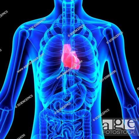
Table of Contents
The Heart: A Hollow Muscular Organ – A Deep Dive into Anatomy, Physiology, and Pathology
The human heart, a fist-sized marvel of biological engineering, is often described simply as a pump. While this is a functional oversimplification, it captures the essence of its primary role: to propel blood throughout the body, delivering oxygen and nutrients while removing waste products. But to truly appreciate the heart's complexity, we must delve deeper into its structure and function, understanding its unique characteristics as a hollow muscular organ.
The Heart's Hollow Structure: Chambers and Valves
The heart's hollow nature is crucial to its function. It isn't a solid mass of muscle, but rather a series of interconnected chambers separated by valves, ensuring unidirectional blood flow. These chambers are:
The Atria: Receiving Chambers
- Right Atrium: Receives deoxygenated blood returning from the body through the superior and inferior vena cava. This blood is low in oxygen and high in carbon dioxide.
- Left Atrium: Receives oxygenated blood from the lungs via the pulmonary veins. This blood is rich in oxygen and low in carbon dioxide.
The atria are relatively thin-walled chambers, acting as receiving reservoirs before blood is propelled into the ventricles.
The Ventricles: Pumping Chambers
- Right Ventricle: Receives deoxygenated blood from the right atrium and pumps it into the pulmonary artery, leading to the lungs for oxygenation. The right ventricle has a thinner muscular wall compared to its left counterpart.
- Left Ventricle: Receives oxygenated blood from the left atrium and pumps it into the aorta, the body's largest artery, distributing oxygenated blood throughout the systemic circulation. The left ventricle has a significantly thicker muscular wall due to the higher pressure required to pump blood throughout the entire body.
The ventricles are the powerful pumping chambers of the heart, responsible for generating the pressure needed to circulate blood. Their muscular walls, composed of cardiac muscle (myocardium), contract forcefully to propel blood forward.
The Valves: Ensuring Unidirectional Flow
The heart valves are critical components, ensuring blood flows in only one direction, preventing backflow and maintaining efficient circulation. There are four key valves:
- Tricuspid Valve: Located between the right atrium and right ventricle.
- Pulmonary Valve: Located at the exit of the right ventricle, leading to the pulmonary artery.
- Mitral (Bicuspid) Valve: Located between the left atrium and left ventricle.
- Aortic Valve: Located at the exit of the left ventricle, leading to the aorta.
These valves open and close passively in response to pressure differences, preventing regurgitation of blood. Their proper function is paramount for maintaining circulatory integrity.
The Myocardium: The Heart's Muscular Engine
The myocardium, the thick muscular layer of the heart, is composed of specialized cardiac muscle cells. These cells are interconnected by intercalated discs, allowing for rapid and synchronized contraction. This synchronized contraction, essential for efficient pumping, is regulated by the heart's intrinsic conduction system.
Cardiac Muscle Cells: Unique Properties
Unlike skeletal muscle cells, cardiac muscle cells possess several unique properties:
- Automaticity: The ability to generate their own electrical impulses, initiating the heartbeat without external stimulation. This is primarily controlled by the sinoatrial (SA) node, the heart's natural pacemaker.
- Excitability: The ability to respond to electrical stimuli, resulting in contraction.
- Conductivity: The ability to transmit electrical impulses rapidly and efficiently throughout the heart.
- Contractility: The ability to contract forcefully, propelling blood throughout the circulatory system.
These properties ensure rhythmic and coordinated contractions, essential for maintaining consistent blood flow.
The Heart's Conduction System: Orchestrating the Beat
The heart's conduction system, a network of specialized cells, ensures the coordinated contraction of the atria and ventricles. This system includes:
- Sinoatrial (SA) Node: The heart's primary pacemaker, initiating the electrical impulse that triggers each heartbeat.
- Atrioventricular (AV) Node: Delays the electrical impulse, allowing the atria to fully contract before the ventricles.
- Bundle of His: Transmits the impulse from the AV node to the ventricles.
- Purkinje Fibers: Distribute the impulse throughout the ventricular myocardium, ensuring coordinated ventricular contraction.
Any disruption in this intricate conduction system can lead to abnormal heart rhythms (arrhythmias).
The Pericardium: Protecting the Heart
The heart isn't simply a free-floating muscular organ; it's enclosed within a protective sac called the pericardium. This fibrous sac has two layers:
- Fibrous Pericardium: The outer layer, a tough, inelastic sac that protects the heart and anchors it to surrounding structures.
- Serous Pericardium: The inner layer, a thin, double-layered membrane with a lubricating fluid (pericardial fluid) between the layers. This fluid minimizes friction during heart contractions.
Inflammation of the pericardium (pericarditis) can cause chest pain and impair heart function. Excessive fluid accumulation in the pericardial space (pericardial effusion) can also compress the heart, reducing its ability to pump effectively (cardiac tamponade).
The Coronary Circulation: Nourishing the Heart Muscle
Even though the heart constantly pumps blood, it doesn't receive its oxygen and nutrients directly from the blood it pumps. Instead, it has its own dedicated circulatory system: the coronary circulation. The coronary arteries branch off from the aorta and supply oxygenated blood to the myocardium. The coronary veins then collect deoxygenated blood and return it to the right atrium.
Blockages in the coronary arteries (coronary artery disease) can significantly reduce blood flow to the heart muscle, leading to myocardial ischemia (lack of oxygen) and potentially a heart attack (myocardial infarction).
Heart Function and Regulation: A Complex Interplay
The heart's function isn't simply a mechanical process; it's tightly regulated by a complex interplay of neural, hormonal, and local factors.
Neural Regulation: The Autonomic Nervous System
The autonomic nervous system, the part of the nervous system that controls involuntary functions, plays a crucial role in regulating heart rate and contractility.
- Sympathetic Nervous System: Increases heart rate and contractility, preparing the body for "fight or flight" responses. This is mediated by norepinephrine.
- Parasympathetic Nervous System: Decreases heart rate and contractility, promoting rest and relaxation. This is mediated by acetylcholine.
Hormonal Regulation: Influencing Heart Function
Several hormones influence heart function, including:
- Epinephrine and Norepinephrine: Released by the adrenal medulla, these hormones mimic the effects of the sympathetic nervous system, increasing heart rate and contractility.
- Thyroid Hormones: Influence heart rate and contractility; high levels can lead to an increased heart rate.
Local Factors: Affecting Heart Contractility
Local factors within the heart muscle itself also play a role in regulating contractility, including:
- Stretch: Increased filling of the heart chambers (preload) stretches the cardiac muscle cells, leading to a more forceful contraction (Frank-Starling mechanism).
- Afterload: The pressure the heart must overcome to eject blood into the arteries. Increased afterload can reduce the efficiency of heart contraction.
Heart Pathology: Common Diseases and Conditions
The heart, despite its remarkable resilience, is susceptible to a range of diseases and conditions. Some of the most common include:
- Coronary Artery Disease (CAD): Atherosclerosis, the buildup of plaque in the coronary arteries, is the primary cause of CAD, leading to reduced blood flow to the heart muscle.
- Heart Failure: The heart's inability to pump enough blood to meet the body's needs. This can result from various underlying conditions.
- Arrhythmias: Irregular heartbeats, caused by disturbances in the heart's conduction system.
- Valvular Heart Disease: Conditions affecting the heart valves, impairing their ability to open and close properly.
- Congenital Heart Defects: Heart abnormalities present at birth.
- Cardiomyopathies: Diseases of the heart muscle itself.
Understanding the heart as a hollow muscular organ allows for a deeper appreciation of its intricate structure and complex physiology. The interplay between its chambers, valves, myocardium, and conduction system, along with the regulatory mechanisms governing its function, is a testament to the wonders of biological engineering. Recognizing the potential for pathology underscores the importance of maintaining cardiovascular health through lifestyle choices and medical attention when necessary. This detailed exploration serves as a foundation for understanding the heart’s crucial role in maintaining overall health and well-being.
Latest Posts
Latest Posts
-
What Is The Lowest Part Of A Wave Called
Apr 07, 2025
-
Are Lysosomes Found In Prokaryotic Cells
Apr 07, 2025
-
7y 4 Y 2 4 3
Apr 07, 2025
-
Lead Nitrate Potassium Iodide Balanced Equation
Apr 07, 2025
-
5 Km Equals How Many Meters
Apr 07, 2025
Related Post
Thank you for visiting our website which covers about The Heart Is A Hollow Muscular Organ . We hope the information provided has been useful to you. Feel free to contact us if you have any questions or need further assistance. See you next time and don't miss to bookmark.
