The Functional Unit Of Skeletal Muscle Is
News Leon
Mar 30, 2025 · 7 min read
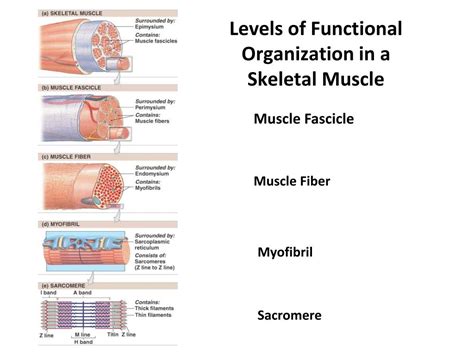
Table of Contents
The Functional Unit of Skeletal Muscle Is: A Deep Dive into the Sarcomere
The human body is a marvel of engineering, capable of incredible feats of strength, endurance, and precision. At the heart of this capacity lies the skeletal muscle system, responsible for locomotion, posture maintenance, and countless other essential functions. But what is the fundamental unit that allows this complex system to function? The answer is the sarcomere. This article will explore the sarcomere in detail, examining its structure, function, and the intricate interplay of proteins that make it the powerhouse of skeletal muscle.
Understanding the Hierarchy of Skeletal Muscle Organization
Before delving into the intricacies of the sarcomere, it's crucial to understand its place within the broader hierarchy of skeletal muscle organization. Skeletal muscle is composed of several nested structures:
1. Muscle: The Whole Organ
This is the macroscopic structure you see and feel – the biceps, triceps, quadriceps, etc. It's a complete organ composed of many bundles of muscle fibers.
2. Muscle Fascicles: Bundles of Muscle Fibers
The muscle is further divided into fascicles, which are bundles of individual muscle fibers held together by connective tissue.
3. Muscle Fibers: Multinucleated Cells
Muscle fibers are elongated, cylindrical cells, also known as muscle cells. Unlike most cells, they are multinucleated, meaning they contain multiple nuclei.
4. Myofibrils: Cylindrical Structures within Muscle Fibers
Each muscle fiber contains numerous myofibrils, cylindrical structures running parallel to the long axis of the fiber. These are the contractile elements of the muscle fiber.
5. Sarcomeres: The Contractile Units of Myofibrils
Finally, we reach the sarcomere, the fundamental unit of contraction within the myofibril. It's the repeating structural and functional unit responsible for muscle contraction. Thousands of sarcomeres arranged end-to-end within each myofibril give the myofibril its characteristic striated appearance under a microscope.
The Sarcomere: Structure and Composition
The sarcomere is defined by the boundaries of two Z-lines (or Z-discs), which are protein structures that act as anchors for the contractile proteins. Within the sarcomere, we find the following key components:
1. Thin Filaments (Actin Filaments):
These filaments are primarily composed of the protein actin, along with other associated proteins like tropomyosin and troponin. Tropomyosin wraps around the actin filament, while troponin is a complex of three proteins that plays a crucial role in regulating muscle contraction. Thin filaments are anchored to the Z-lines.
2. Thick Filaments (Myosin Filaments):
These filaments are mainly composed of the protein myosin. Each myosin molecule has a head and a tail. The myosin heads are crucial for the interaction with actin during muscle contraction. Thick filaments are located in the center of the sarcomere, overlapping with the thin filaments.
3. A-Band: The Dark Band
This is the region of the sarcomere where thick filaments are present, including the areas where thick and thin filaments overlap. It appears dark under a microscope due to the dense packing of these filaments.
4. I-Band: The Light Band
This is the region of the sarcomere containing only thin filaments. It appears light under a microscope because it lacks the dense packing of the thick filaments.
5. H-Zone: The Lighter Region within the A-Band
This is the central region of the A-band where only thick filaments are present, without overlap from thin filaments.
6. M-Line: The Middle of the Sarcomere
This is the structural protein complex located in the center of the H-zone. It helps to hold the thick filaments in place and maintain the structural integrity of the sarcomere.
The Sliding Filament Theory: How Sarcomeres Contract
The mechanism by which sarcomeres contract is elegantly explained by the sliding filament theory. This theory states that muscle contraction occurs due to the sliding of thin filaments past thick filaments, resulting in a shortening of the sarcomere and, consequently, the entire muscle fiber.
This process is driven by the interaction between myosin heads and actin filaments. The myosin heads bind to specific sites on the actin filament, forming cross-bridges. The myosin heads then undergo a conformational change, pulling the thin filaments towards the center of the sarcomere. This process is fueled by the hydrolysis of ATP (adenosine triphosphate), the energy currency of cells. The cycle of cross-bridge formation, power stroke, detachment, and resetting repeats multiple times, leading to the progressive shortening of the sarcomere.
Key steps in the cross-bridge cycle:
- ATP binding: Myosin head binds to ATP, causing detachment from actin.
- ATP hydrolysis: ATP is hydrolyzed to ADP and Pi (inorganic phosphate), causing a conformational change in the myosin head.
- Cross-bridge formation: The energized myosin head binds to actin.
- Power stroke: Pi is released, causing the myosin head to pivot and pull the thin filament towards the center of the sarcomere.
- ADP release: ADP is released, maintaining the strong bond between myosin and actin.
- ATP binding (repeat): A new ATP molecule binds to the myosin head, causing detachment from actin and the cycle begins anew.
Regulation of Muscle Contraction: The Role of Calcium Ions
The process of muscle contraction is tightly regulated to ensure precise control over muscle force and movement. Calcium ions (Ca2+) play a central role in this regulation.
When a nerve impulse reaches a muscle fiber, it triggers the release of Ca2+ from the sarcoplasmic reticulum (SR), a specialized intracellular organelle that stores Ca2+. The released Ca2+ binds to troponin, causing a conformational change in the troponin-tropomyosin complex. This change exposes the myosin-binding sites on the actin filament, allowing the myosin heads to bind and initiate the cross-bridge cycle.
When the nerve impulse ceases, Ca2+ is actively pumped back into the SR, removing it from the myofilaments. This causes the troponin-tropomyosin complex to return to its original conformation, blocking the myosin-binding sites and preventing further contraction. This precise control of Ca2+ levels is essential for the coordinated and controlled movements of our muscles.
Types of Muscle Contractions: Isometric and Isotonic
Sarcomeres and the sliding filament mechanism allow for two main types of muscle contractions:
1. Isometric Contractions:
In isometric contractions, the muscle generates force without changing its length. This occurs when the muscle attempts to move a load that is too heavy to lift. The sarcomeres generate force, but the overall muscle length remains constant because the external load is greater than the force the muscle produces. Think of trying to push a wall – your muscles generate force but don’t shorten.
2. Isotonic Contractions:
In isotonic contractions, the muscle generates force and changes its length. This is the type of contraction that most commonly occurs during movement. There are two subtypes:
- Concentric contractions: The muscle shortens while generating force. For example, bending your elbow to lift a weight.
- Eccentric contractions: The muscle lengthens while generating force. For example, slowly lowering a weight after you've lifted it.
Sarcomere Dysfunction and Muscle Diseases
Proper sarcomere function is essential for healthy muscle performance. Disruptions in the structure or function of sarcomeric proteins can lead to various muscle diseases, including:
- Muscular dystrophies: A group of inherited diseases characterized by progressive muscle weakness and degeneration. Mutations in genes encoding sarcomeric proteins, such as dystrophin, are often the cause.
- Cardiomyopathies: Diseases affecting the heart muscle, often involving defects in sarcomeric proteins.
- Myopathies: A broad category of muscle diseases that can involve various causes, including genetic mutations, infections, and autoimmune disorders.
Conclusion: The Sarcomere – A Microscopic Masterpiece
The sarcomere, though microscopic in size, is a masterpiece of biological engineering. Its elegant structure and the precise interplay of its protein components allow for the generation of force and movement that are essential for life. Understanding the sarcomere provides a crucial foundation for comprehending the intricacies of muscle function, as well as the mechanisms underlying various muscle diseases. Future research focused on the sarcomere holds immense potential for developing new treatments and therapies for these debilitating conditions. The continued exploration of this fundamental unit will undoubtedly reveal further insights into the complexity and remarkable capabilities of the human musculoskeletal system.
Latest Posts
Latest Posts
-
Number Of Chromosomes In A Fruit Fly
Apr 01, 2025
-
Two Lines Perpendicular To The Same Plane Are
Apr 01, 2025
-
How To Find The Moles Of An Element
Apr 01, 2025
-
What Did David Used To Kill Goliath
Apr 01, 2025
-
Are Cells Depicted Plant Or Animal
Apr 01, 2025
Related Post
Thank you for visiting our website which covers about The Functional Unit Of Skeletal Muscle Is . We hope the information provided has been useful to you. Feel free to contact us if you have any questions or need further assistance. See you next time and don't miss to bookmark.
