The First Heart Sound Is The Closing Of The
News Leon
Mar 21, 2025 · 6 min read
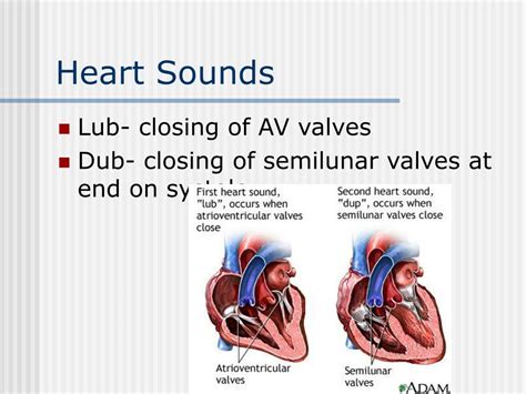
Table of Contents
The First Heart Sound: The Closing of the Atrioventricular Valves
The rhythmic lub-dub of the heartbeat is a familiar sound, a constant companion throughout our lives. But what actually creates these sounds? Understanding the mechanics behind the heart sounds, particularly the "lub," is crucial to comprehending cardiovascular health and disease. This article delves deep into the physiology of the first heart sound (S1), focusing on its primary source: the closing of the atrioventricular (AV) valves.
The Cardiac Cycle and Valve Function: Setting the Stage
Before exploring the intricacies of S1, we need to establish a solid foundation in the cardiac cycle and the roles of the heart valves. The heart, a tireless pump, propels blood through a continuous circuit. This cyclical process involves coordinated contractions and relaxations of the atria and ventricles, regulated by electrical impulses. The four heart valves—the mitral and tricuspid valves (AV valves) and the aortic and pulmonic valves (semilunar valves)—ensure unidirectional blood flow.
Atrioventricular Valves: Guardians of the Ventricles
The AV valves, positioned between the atria and ventricles, prevent backflow of blood from the ventricles into the atria during ventricular contraction (systole). The mitral valve, located between the left atrium and left ventricle, has two cusps (leaflets). The tricuspid valve, situated between the right atrium and right ventricle, possesses three cusps. These valves are anchored by strong chordae tendineae, which connect to papillary muscles in the ventricular walls. These structures play a crucial role in preventing valve prolapse during ventricular contraction.
Semilunar Valves: Controlling Arterial Flow
The semilunar valves, the aortic and pulmonic valves, are positioned at the exits of the ventricles. They prevent backflow of blood from the aorta and pulmonary artery into the ventricles during ventricular relaxation (diastole). Unlike the AV valves, the semilunar valves don't have chordae tendineae; their structure relies on the pressure differences between the ventricles and the great vessels.
The Genesis of the First Heart Sound (S1): A Closer Look
The first heart sound, S1, is a relatively low-pitched, dull sound heard at the beginning of systole. Its primary acoustic source is the closure of the mitral and tricuspid valves. As the ventricles contract, ventricular pressure surpasses atrial pressure, forcing the AV valves to close. This closure is a complex mechanical event that produces vibrations that are transmitted to the chest wall, creating the audible S1.
Factors Influencing S1 Intensity
The intensity of S1 is not uniform across all heartbeats; several factors can influence its loudness and character:
- Rate of ventricular contraction: A faster ventricular contraction leads to a more forceful valve closure and a louder S1. Conversely, a slower contraction results in a softer S1.
- Valve leaflet thickness and mobility: Thickened or stiff valve leaflets, often seen in conditions like calcific mitral stenosis, result in a louder S1. Conversely, impaired leaflet mobility, as in mitral valve prolapse, can lead to a softer or split S1.
- Preload and afterload: Increased preload (the amount of blood in the ventricles before contraction) and afterload (the resistance the ventricle must overcome to eject blood) can influence the force of valve closure and, consequently, S1 intensity.
- Position of the heart: The anatomical position of the heart and the patient's body position can affect the transmission of sound to the chest wall, impacting the perceived loudness of S1.
Splitting of S1: A Normal Variation or a Sign of Pathology?
While S1 is typically heard as a single sound, it can sometimes be perceived as split, meaning two distinct components are audible. This splitting is usually physiological and is caused by a slight temporal discrepancy between the closure of the mitral and tricuspid valves. The mitral valve usually closes slightly before the tricuspid valve, but this timing difference is often too subtle to be heard. However, certain conditions like right bundle branch block or atrial septal defect can exaggerate this timing difference, resulting in a more audible split S1.
Differentiating S1 from Other Heart Sounds: Avoiding Confusion
Accurate interpretation of heart sounds requires careful differentiation between S1 and other potential cardiac sounds. Mistaking S1 for other sounds can lead to misdiagnosis and inappropriate management.
S1 vs. S2: Distinguishing the Two Major Sounds
S2, the second heart sound, results from the closure of the semilunar valves (aortic and pulmonic). It's generally higher pitched and shorter than S1. The key difference lies in the timing relative to the cardiac cycle. S1 marks the beginning of systole, while S2 marks the end.
S1 vs. Extra Heart Sounds: Identifying Additional Sounds
Extra heart sounds, such as S3 and S4, can be mistaken for S1 if not carefully examined. S3, a low-pitched sound heard in early diastole, often reflects rapid ventricular filling. S4, another low-pitched sound heard in late diastole, is associated with atrial contraction against a stiff or hypertrophic ventricle. These extra sounds are often associated with specific conditions and require further investigation.
Clinical Significance of S1: Diagnostic Implications
Assessment of S1 is a crucial part of the physical examination of the cardiovascular system. Changes in the timing, intensity, and character of S1 can provide valuable clues to underlying cardiac pathology.
Altered S1 Intensity: Indicators of Disease
A loud S1 can be indicative of conditions causing increased ventricular contractility or stiff AV valves, such as:
- Mitral stenosis: Narrowing of the mitral valve orifice increases pressure and flow, leading to a louder S1.
- Hypertrophic cardiomyopathy: Increased ventricular muscle mass results in enhanced contractility and a louder S1.
A soft or absent S1 can suggest:
- Mitral regurgitation: Backflow of blood from the left ventricle to the left atrium reduces the effective closure of the mitral valve.
- First-degree heart block: Prolonged AV nodal conduction slows ventricular activation, delaying S1.
Variations in S1 Timing: Clues to Conduction Disorders
Changes in the timing of S1 can provide information about conduction abnormalities within the heart. For example, a prolonged PR interval on the electrocardiogram (ECG), indicative of AV conduction delays, can be associated with a delayed S1.
Auscultation Techniques for Assessing S1: Refining Your Skills
Accurate assessment of S1 requires a systematic approach to auscultation. Using the diaphragm and bell of the stethoscope, careful listening at various locations on the chest wall helps to determine the character and timing of S1. Comparing the intensity of S1 between different auscultation points can further refine the diagnosis.
Conclusion: The Significance of S1 in Cardiovascular Assessment
The first heart sound, S1, arising primarily from the closure of the atrioventricular valves, is a cornerstone of cardiovascular assessment. Understanding its physiology, influencing factors, and diagnostic implications is paramount for healthcare professionals. Changes in the timing, intensity, and character of S1 can indicate underlying cardiac pathology, highlighting the importance of meticulous auscultation and integration of other diagnostic modalities for accurate diagnosis and appropriate management of cardiovascular disease. The seemingly simple lub of the heartbeat contains a wealth of information, unlocking insights into the intricate workings of the human heart.
Latest Posts
Latest Posts
-
A Rotating Fan Completes 1200 Revolutions
Mar 28, 2025
-
1 Meter Equals How Many Millimeters
Mar 28, 2025
-
In The System Of Mass Production Unskilled Workers
Mar 28, 2025
-
What Is The Ph Of The Neutral Solution
Mar 28, 2025
-
Respiratory Control Centers Are Located In The
Mar 28, 2025
Related Post
Thank you for visiting our website which covers about The First Heart Sound Is The Closing Of The . We hope the information provided has been useful to you. Feel free to contact us if you have any questions or need further assistance. See you next time and don't miss to bookmark.
