Structure From Which Chordae Tendineae Originate
News Leon
Mar 15, 2025 · 7 min read
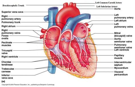
Table of Contents
Structure From Which Chordae Tendineae Originate: A Comprehensive Guide
The heart, a tireless engine driving life's processes, is a marvel of intricate engineering. Within its chambers, a delicate yet crucial interplay of structures ensures the efficient pumping of blood. Central to this intricate system are the chordae tendineae, thin, strong, tendon-like cords that play a vital role in preventing valve prolapse during ventricular contraction. Understanding the precise structure from which these chordae tendineae originate is crucial for comprehending the mechanics of the heart and diagnosing associated pathologies. This article will delve deep into this fascinating anatomical aspect, exploring the origins, attachments, and functional significance of the chordae tendineae.
The Papillary Muscles: The Primary Origin of Chordae Tendineae
The primary origin of the chordae tendineae is the papillary muscles. These are cone-shaped muscular projections located within the ventricles, the heart's lower chambers. Their strategic placement is critical to their function. They are not directly attached to the ventricular walls but arise from the trabeculae carneae, the irregular muscular ridges lining the ventricular surfaces. This indirect attachment allows for a degree of flexibility and movement that is essential for efficient valve function.
Number and Distribution of Papillary Muscles
The number and arrangement of papillary muscles vary slightly between individuals, but a general pattern exists. The right ventricle, typically possessing fewer and less robust papillary muscles, often features three, though variations can occur. The left ventricle, on the other hand, generally presents two prominent papillary muscles – an anterior and a posterior – although additional smaller muscles can be observed. This difference in the number and arrangement reflects the differing hemodynamic pressures experienced by the two ventricles. The left ventricle, responsible for pumping oxygenated blood to the systemic circulation, faces significantly higher pressures than the right ventricle, requiring a more robust structural support system for the atrioventricular valves.
Microscopic Anatomy of Papillary Muscles
At a microscopic level, papillary muscles consist of cardiac muscle fibers arranged in a complex interwoven pattern. These fibers are richly supplied with capillaries and nerves, reflecting the muscles' high metabolic demands. The arrangement of these fibers contributes to the papillary muscle's ability to generate the force needed to anchor the chordae tendineae and maintain valve integrity. The precise organization of these muscle fibers is still a subject of ongoing research, with studies focusing on the relationship between fiber orientation and contractile function.
Attachment Points and Functional Implications
The chordae tendineae, originating from the papillary muscles, fan out to attach to the cusps (leaflets) of the atrioventricular valves. These valves – the tricuspid valve on the right side and the mitral valve on the left – control the unidirectional flow of blood from the atria to the ventricles. Each cusp receives multiple chordae tendineae from different papillary muscles, creating a complex network of attachments. This arrangement provides redundancy, ensuring that even if one chordae tendineae fails, the valve's integrity is maintained.
Understanding the Complex Network: Redundancy and Robustness
The intricate arrangement of chordae tendineae ensures robust valve function. Several chordae tendineae attach to each cusp, preventing a single point of failure. This arrangement is crucial in managing the substantial pressure changes that occur during the cardiac cycle. If a single chordae tendineae ruptures, other supporting chords can compensate, preventing catastrophic valve dysfunction. This inherent redundancy highlights the sophistication of the cardiovascular system's design.
Variations in Attachment Patterns and their Significance
While a general pattern exists, individual variations in the number and attachment points of chordae tendineae are commonly observed. These variations don’t necessarily imply pathology, but understanding these anatomical nuances is vital for surgeons and cardiologists. Variations in the number and position of chordae tendineae can impact surgical approaches during procedures involving the atrioventricular valves, emphasizing the need for meticulous anatomical assessment before any intervention.
Functional Significance of the Chordae Tendineae Origin
The strategic origin of the chordae tendineae from the papillary muscles is directly linked to their crucial function in preventing valve prolapse. During ventricular contraction, the pressure within the ventricles rises dramatically. Without the chordae tendineae, this increase in pressure would force the atrioventricular valves open in the wrong direction, leading to backflow of blood into the atria (regurgitation). The papillary muscles contract synchronously with the ventricles, tightening the chordae tendineae and pulling the valve cusps closed, preventing this potentially devastating backward flow.
Coordination and Timing: A Symphony of Contraction
The precise coordination between papillary muscle contraction and ventricular contraction is crucial for efficient heart function. Any disruption in this finely tuned synchronization can lead to valve dysfunction. The timing of papillary muscle contraction is tightly regulated by the cardiac conduction system, ensuring the valves close at the appropriate moment during the cardiac cycle. Studies investigating the precise electrophysiological mechanisms controlling papillary muscle activation are ongoing and contribute to a deeper understanding of heart function.
Impact of Papillary Muscle Dysfunction
Dysfunction of the papillary muscles, whether due to ischemia, infarction, or other pathologies, can significantly impair the function of the atrioventricular valves. This can manifest as mitral regurgitation or tricuspid regurgitation, leading to a range of symptoms, including shortness of breath, fatigue, and edema. Understanding the origin and function of the chordae tendineae is paramount in diagnosing and managing these conditions.
Clinical Significance and Related Pathologies
The structural integrity of the papillary muscles and their attachments to the chordae tendineae are vital for maintaining normal cardiac function. Several clinical conditions can affect this delicate system, highlighting the importance of comprehending its anatomical underpinnings.
Papillary Muscle Rupture
Papillary muscle rupture is a serious complication that can occur following myocardial infarction (heart attack). Damage to the papillary muscle can lead to its rupture, resulting in severe mitral regurgitation or tricuspid regurgitation. This condition requires urgent medical intervention, often involving surgery to repair or replace the damaged valve. The location and extent of the rupture significantly influence the severity of the regurgitation and the chosen treatment strategy.
Chordae Tendineae Rupture
Chordae tendineae rupture can occur independently of papillary muscle dysfunction, often due to degenerative processes or trauma. This rupture can also lead to significant valve regurgitation, necessitating medical intervention. The rupture’s location and the number of affected chordae tendineae influence the severity of the resulting valve dysfunction and the treatment approach.
Myocardial Infarction and Valve Dysfunction
Myocardial infarction can indirectly affect the papillary muscles and chordae tendineae, leading to ventricular dysfunction and subsequent valve complications. Ischemia (reduced blood flow) to the papillary muscles can impair their contractility, diminishing their ability to support the atrioventricular valves. This can result in valve prolapse and regurgitation.
Advanced Imaging Techniques and Diagnosis
Modern medical imaging techniques play a crucial role in diagnosing pathologies related to the papillary muscles and chordae tendineae.
Echocardiography
Echocardiography, particularly transthoracic echocardiography (TTE) and transesophageal echocardiography (TEE), provides detailed images of the heart’s structures, including the papillary muscles and chordae tendineae. These techniques allow clinicians to assess valve function, identify areas of rupture or dysfunction, and guide treatment decisions. The high resolution and ability to visualize the heart in real-time make echocardiography an invaluable diagnostic tool.
Cardiac Magnetic Resonance Imaging (CMRI)
CMRI offers high-resolution images of the heart and provides detailed anatomical information, enabling precise assessment of papillary muscle size, shape, and function. CMRI can be particularly useful in detecting subtle abnormalities that may be missed by other imaging modalities. Its superior soft tissue contrast enhances the visualization of the papillary muscles and their attachments.
Conclusion: The Importance of Understanding the Chordae Tendineae Origin
The structure from which chordae tendineae originate – the papillary muscles – plays a pivotal role in maintaining normal heart valve function. The intricate interplay between papillary muscles, chordae tendineae, and atrioventricular valves ensures efficient blood flow through the heart. Understanding the anatomy, function, and clinical significance of this complex system is essential for clinicians in diagnosing and managing a range of cardiac pathologies. Further research into the subtle variations in structure and function, as well as the impact of various pathologies, will continue to refine our understanding of this vital aspect of cardiovascular health. The ongoing advancements in medical imaging techniques allow for more precise diagnoses and individualized treatment strategies, significantly improving patient outcomes. The continued exploration of the complex interplay of these structures will undoubtedly lead to further breakthroughs in the field of cardiology.
Latest Posts
Latest Posts
-
The Old Man And The Sea Plot Summary
Mar 15, 2025
-
How Many Minutes Are There In One Day
Mar 15, 2025
-
A Sphere Has How Many Vertex
Mar 15, 2025
-
What Is The Electron Configuration For Ne
Mar 15, 2025
-
Tool Used To Detect Electric Charge
Mar 15, 2025
Related Post
Thank you for visiting our website which covers about Structure From Which Chordae Tendineae Originate . We hope the information provided has been useful to you. Feel free to contact us if you have any questions or need further assistance. See you next time and don't miss to bookmark.
