Prevents Backflow Into The Right Atrium
News Leon
Mar 25, 2025 · 7 min read
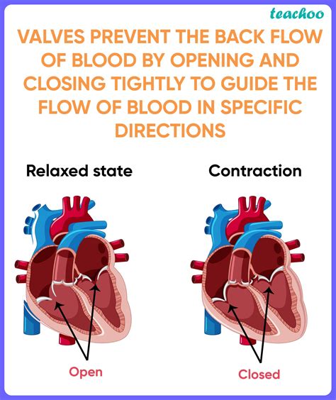
Table of Contents
Preventing Backflow into the Right Atrium: A Comprehensive Guide
The heart, a marvel of biological engineering, relies on a precise system of valves to ensure unidirectional blood flow. Backflow, or regurgitation, can severely compromise this efficiency, leading to various cardiovascular complications. One crucial area where backflow must be prevented is the flow from the right ventricle into the right atrium. This article delves into the mechanisms preventing this backflow, the consequences of its failure, and potential associated conditions.
Understanding the Tricupsid Valve and its Role
The primary mechanism preventing backflow from the right ventricle into the right atrium is the tricuspid valve. This valve, comprised of three cusps or leaflets (hence "tri-cuspid"), acts as a one-way door, allowing oxygen-poor blood to flow from the right atrium to the right ventricle during ventricular diastole (relaxation) but preventing its return during ventricular systole (contraction).
The Structure of the Tricupsid Valve
The tricuspid valve's structure is intricately designed to facilitate its function. Each cusp is composed of fibrous tissue covered by endocardium, the inner lining of the heart. These cusps are attached to papillary muscles within the right ventricle via chordae tendineae, strong fibrous cords resembling tiny strings.
- Papillary Muscles: These muscles contract during ventricular systole, preventing the cusps from inverting (prolapsing) into the right atrium under the pressure of blood being ejected into the pulmonary artery. Their coordinated action is crucial for maintaining valve integrity.
- Chordae Tendineae: These “heart strings” act as a supportive framework, connecting the papillary muscles to the valve cusps. They prevent excessive stretching and prolapse of the cusps, thus ensuring proper valve closure.
- Annulus Fibrosus: The tricuspid valve is anchored to the fibrous ring (annulus fibrosus) surrounding the atrioventricular orifice. This provides a stable base for the valve's function and prevents displacement.
The Mechanism of Tricuspid Valve Closure
The intricate interplay between the papillary muscles, chordae tendineae, and valve cusps orchestrates the precise closure of the tricuspid valve. During ventricular systole, as the right ventricle contracts, pressure within the ventricle rises significantly. This pressure pushes the tricuspid valve cusps together, effectively sealing the opening between the right atrium and ventricle. Simultaneously, the papillary muscles contract, tightening the chordae tendineae and further reinforcing the closure. This prevents backflow of blood into the right atrium.
Consequences of Tricuspid Regurgitation
When the tricuspid valve fails to close properly, allowing blood to flow back into the right atrium during ventricular systole, the condition is known as tricuspid regurgitation (TR). This backflow reduces the effectiveness of the heart's pumping action, leading to a range of symptoms and complications:
Reduced Cardiac Output
The primary consequence of TR is reduced cardiac output. The backflow of blood from the right ventricle to the right atrium effectively reduces the volume of blood ejected into the pulmonary artery with each heartbeat. This decreased blood flow to the lungs can lead to inadequate oxygenation of the blood.
Right Atrial Enlargement
The continuous backflow into the right atrium puts added pressure on this chamber, causing it to enlarge over time. This enlargement, known as right atrial dilation, can further compromise its function and potentially lead to atrial fibrillation, an irregular heartbeat.
Right Ventricular Hypertrophy
The right ventricle works harder to compensate for the backflow, leading to right ventricular hypertrophy. This means the muscle of the right ventricle thickens in an attempt to maintain adequate cardiac output. While initially beneficial, hypertrophy can eventually impair the ventricle's ability to function effectively.
Peripheral Edema
Reduced cardiac output due to TR can lead to fluid buildup in the peripheral tissues, causing edema, particularly in the legs and ankles. This fluid accumulation is a consequence of the heart's inability to effectively pump blood throughout the circulatory system.
Liver Congestion
The increased pressure in the right atrium can also lead to liver congestion (hepatomegaly). This is because the inferior vena cava, which returns blood from the lower body, including the liver, drains into the right atrium. Increased pressure can impede the flow of blood out of the liver.
Symptoms of Tricuspid Regurgitation
The symptoms of TR can vary depending on the severity of the regurgitation and the individual's overall health. Some common symptoms include:
- Fatigue: Due to the reduced cardiac output and inadequate oxygenation.
- Shortness of breath (dyspnea): Especially during exertion.
- Edema (swelling): In the legs, ankles, and feet.
- Jugular venous distension (JVD): Visible bulging of the neck veins.
- Abdominal distension: Due to liver congestion.
- Murmur: An abnormal heart sound heard during a physical examination.
Causes of Tricuspid Regurgitation
Tricuspid regurgitation can arise from various underlying conditions, including:
Congenital Heart Defects
Some individuals are born with defects in the tricuspid valve, such as a malformed valve or abnormal chordae tendineae. These defects can lead to TR from an early age.
Rheumatic Fever
Rheumatic fever, a complication of untreated streptococcal infections, can damage the heart valves, including the tricuspid valve, leading to TR.
Infective Endocarditis
Infection of the heart valves (infective endocarditis) can weaken the tricuspid valve and impair its ability to close properly.
Cardiomyopathies
Diseases that affect the heart muscle (cardiomyopathies) can weaken the papillary muscles or the right ventricle, contributing to TR.
Pulmonary Hypertension
Increased pressure in the pulmonary artery (pulmonary hypertension) places extra stress on the tricuspid valve, leading to its dysfunction and regurgitation.
Diagnosis of Tricuspid Regurgitation
Diagnosing TR involves a comprehensive evaluation combining several methods:
Physical Examination
The doctor will listen to the heart using a stethoscope. A characteristic murmur, often described as a systolic murmur, may be heard, indicating backflow. Other signs such as JVD and peripheral edema can also provide clues.
Echocardiography
This non-invasive imaging technique uses ultrasound to visualize the heart's structure and function. Echocardiography allows precise assessment of the tricuspid valve's morphology, degree of regurgitation, and its impact on the right atrium and ventricle. It's the gold standard for diagnosing and assessing TR severity.
Electrocardiogram (ECG)
An ECG records the electrical activity of the heart. While it may not directly visualize the valve, it can reveal signs of right atrial or ventricular enlargement, suggesting the presence of TR.
Cardiac Catheterization
In certain cases, cardiac catheterization may be necessary. This invasive procedure allows direct measurement of pressures within the heart chambers and provides more detailed information about the severity of TR and associated conditions.
Management of Tricuspid Regurgitation
Treatment for TR depends on the severity of the condition and its underlying cause. Mild TR may not require specific treatment beyond regular monitoring. However, more severe cases may require interventions:
Medical Management
Medical management focuses on addressing underlying conditions such as pulmonary hypertension or cardiomyopathies. Diuretics may be used to reduce fluid buildup, while medications to manage heart failure may be necessary.
Surgical Intervention
In cases of severe TR refractory to medical management, surgical intervention may be necessary. Surgical options include:
- Tricuspid Valve Repair: This aims to restore the valve's function by repairing damaged cusps or chordae tendineae. It's often preferred over valve replacement whenever possible.
- Tricuspid Valve Replacement: In cases where repair is not feasible, a damaged tricuspid valve may be replaced with a mechanical or bioprosthetic valve.
Preventing Tricuspid Regurgitation
While not all cases of TR are preventable, certain measures can reduce the risk:
Managing Underlying Conditions
Effective management of conditions such as rheumatic fever, infective endocarditis, and pulmonary hypertension is crucial in preventing or delaying the onset of TR.
Maintaining a Healthy Lifestyle
A healthy lifestyle including regular exercise, a balanced diet, and avoiding smoking can help maintain overall cardiovascular health and reduce the risk of developing heart conditions that may lead to TR.
This comprehensive guide provides a thorough understanding of the mechanisms preventing backflow into the right atrium, the consequences of tricuspid regurgitation, its causes, diagnosis, management, and preventive measures. Understanding this complex interplay is crucial for early detection and effective management of this significant cardiovascular condition. Remember, always consult a healthcare professional for any concerns regarding your heart health. Early diagnosis and appropriate intervention are key to improving outcomes and quality of life for individuals affected by tricuspid regurgitation.
Latest Posts
Latest Posts
-
A Rotating Fan Completes 1200 Revolutions
Mar 28, 2025
-
1 Meter Equals How Many Millimeters
Mar 28, 2025
-
In The System Of Mass Production Unskilled Workers
Mar 28, 2025
-
What Is The Ph Of The Neutral Solution
Mar 28, 2025
-
Respiratory Control Centers Are Located In The
Mar 28, 2025
Related Post
Thank you for visiting our website which covers about Prevents Backflow Into The Right Atrium . We hope the information provided has been useful to you. Feel free to contact us if you have any questions or need further assistance. See you next time and don't miss to bookmark.
