Place The Following Parts Of A Reflex Arc In Order.
News Leon
Mar 25, 2025 · 6 min read
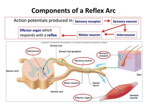
Table of Contents
The Reflex Arc: A Step-by-Step Guide to Understanding Your Body's Automatic Responses
The human body is a marvel of intricate design, capable of incredible feats of coordination and reaction. At the heart of many of these rapid responses lies the reflex arc, a neural pathway that allows for quick, involuntary reactions to stimuli. Understanding the components and sequence of a reflex arc is crucial to grasping the fundamental mechanisms of our nervous system. This comprehensive guide will delve into the intricacies of the reflex arc, explaining each component and placing them in the correct order, accompanied by illustrative examples.
What is a Reflex Arc?
A reflex arc is a neural pathway that controls a reflex action. Reflex actions are rapid, automatic, and involuntary responses to a specific stimulus. They bypass the brain's conscious processing centers, allowing for incredibly fast reactions – crucial for survival in situations requiring immediate action, such as withdrawing your hand from a hot stove or blinking when something approaches your eye. This speed is achieved because the signal processing occurs entirely within the spinal cord or brainstem, rather than traveling all the way to the brain and back.
The Five Essential Components of a Reflex Arc
The reflex arc consists of five key components, arranged in a specific sequence:
-
Receptor: This is the specialized structure that detects the stimulus. Receptors can be simple nerve endings or more complex sensory organs. Examples include:
- Mechanoreceptors: Detect physical pressure, touch, or vibration (e.g., Pacinian corpuscles in the skin).
- Thermoreceptors: Detect changes in temperature (e.g., free nerve endings in the skin).
- Nociceptors: Detect pain (e.g., free nerve endings in the skin and internal organs).
- Photoreceptors: Detect light (e.g., rods and cones in the retina).
-
Sensory Neuron (Afferent Neuron): This neuron transmits the sensory information from the receptor to the central nervous system (CNS), which includes the brain and spinal cord. The sensory neuron's cell body is located in the dorsal root ganglion, a cluster of nerve cell bodies outside the spinal cord. The axon of the sensory neuron carries the signal towards the CNS. This transmission is often described as the afferent pathway.
-
Interneuron (Association Neuron): This neuron acts as a connector or relay within the CNS. Not all reflex arcs involve an interneuron. In simple reflex arcs, the sensory neuron directly synapses with the motor neuron. However, in more complex reflexes, interneurons integrate information from multiple sensory neurons and may connect to other interneurons before finally reaching the motor neuron. The interneuron facilitates complex processing and coordination. The interneuron plays a critical role in allowing the reflex arc to be modified by other neural pathways, for example, allowing for a conscious experience of the stimulus after the reflex has occurred.
-
Motor Neuron (Efferent Neuron): This neuron transmits the impulse from the CNS to the effector organ, triggering the response. The motor neuron's cell body is located within the anterior horn of the spinal cord, and its axon extends to the effector. This transmission is described as the efferent pathway.
-
Effector: This is the muscle or gland that carries out the response. The effector responds to the nerve impulse from the motor neuron and produces a noticeable effect. Examples include:
- Muscles: Responsible for movement, such as contracting to withdraw a limb from a painful stimulus.
- Glands: Responsible for secretion, such as releasing sweat in response to heat.
The Sequence of Events in a Reflex Arc: A Step-by-Step Breakdown
Let's illustrate the sequence with a common example: the knee-jerk reflex.
-
Stimulus: A sharp tap below the kneecap stretches the quadriceps tendon.
-
Receptor: Muscle spindles within the quadriceps muscle detect the stretch. These specialized receptors are sensitive to changes in muscle length.
-
Sensory Neuron: The sensory neuron transmits the signal from the muscle spindle along its axon to the spinal cord. The signal is transmitted as a series of action potentials, electrochemical signals that propagate down the neuron's axon.
-
Interneuron (optional, in this simple reflex): In the knee-jerk reflex, the sensory neuron directly synapses with the motor neuron without an intermediary interneuron. However, in more complex reflexes involving integration or coordination, an interneuron facilitates communication between multiple neurons.
-
Motor Neuron: The motor neuron receives the signal and transmits it along its axon to the effector.
-
Effector: The quadriceps muscle, acting as the effector, contracts, causing the leg to extend. Simultaneously, another motor neuron inhibits the hamstring muscle (antagonist), allowing for smooth extension without resistance. This reciprocal inhibition is crucial for coordinated movement.
-
Response: The leg kicks forward – a classic example of a monosynaptic reflex (a reflex arc involving only one synapse between the sensory and motor neuron).
Examples of Different Reflex Arcs
The knee-jerk reflex is a simple example. More complex reflexes involve multiple synapses and interneurons, allowing for greater integration and control. Examples include:
-
Withdrawal Reflex: Touching a hot stove triggers nociceptors in the skin. The sensory neuron transmits the signal to the spinal cord, where interneurons connect to motor neurons controlling the muscles in the arm and hand. These muscles contract, causing the hand to withdraw rapidly from the heat source. This is a polysynaptic reflex, involving multiple synapses. Often, a simultaneous reflex occurs in the opposite limb, extending it to maintain balance – a demonstration of how interneurons coordinate different muscle groups.
-
Pupillary Light Reflex: Shining a light into the eye causes the pupils to constrict. Photoreceptors in the retina detect the light, sending signals via sensory neurons to the brain stem. Interneurons process the signals, leading to motor neuron activation. The motor neurons innervate the muscles of the iris, resulting in pupil constriction.
-
Gag Reflex: Touching the back of the throat triggers sensory receptors, leading to a coordinated response involving multiple muscles in the throat and mouth region.
Clinical Significance of Reflex Arcs
Assessing reflex arcs is a vital part of neurological examinations. Abnormal reflexes can indicate damage to the nervous system, such as:
-
Hyporeflexia: Diminished or absent reflexes, which might suggest damage to the peripheral nerves or spinal cord.
-
Hyperreflexia: Exaggerated reflexes, indicating possible damage to the upper motor neurons in the brain or spinal cord.
-
Clonus: Rhythmic, involuntary muscle contractions, indicative of certain neurological conditions.
Conclusion: The Reflex Arc – A Foundation of Neurological Function
The reflex arc represents a fundamental principle of neurological function. Its intricate design, involving a specific sequence of interconnected neurons and effector organs, allows for rapid, automatic responses to stimuli that are crucial for survival and everyday actions. Understanding the components and operation of the reflex arc is essential for appreciating the complexity and efficiency of the human nervous system. The speed and precision of these involuntary actions showcase the intricate interplay of receptors, sensory and motor neurons, and effectors working in perfect synchrony, contributing to the overall remarkable functioning of the human body. Further research and understanding of the reflex arc's nuances promise to continue to illuminate the mysteries of neurological functioning and facilitate better treatment of related neurological disorders.
Latest Posts
Latest Posts
-
Round 64 To The Nearest Ten
Mar 26, 2025
-
Which Of The Following Are Meso Compounds
Mar 26, 2025
-
Words Beginning With The Same Letter
Mar 26, 2025
-
The Proper Order For The Scientific Process Is
Mar 26, 2025
-
What Is The Lightest Element On The Periodic Table
Mar 26, 2025
Related Post
Thank you for visiting our website which covers about Place The Following Parts Of A Reflex Arc In Order. . We hope the information provided has been useful to you. Feel free to contact us if you have any questions or need further assistance. See you next time and don't miss to bookmark.
