Optic Nerve And Blood Vessels Enter The Eye At The
News Leon
Mar 18, 2025 · 7 min read
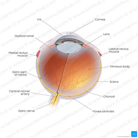
Table of Contents
Optic Nerve and Blood Vessels Enter the Eye at the Optic Disc: A Comprehensive Overview
The human eye, a marvel of biological engineering, is responsible for our sense of sight. Its intricate structure allows us to perceive light, color, and detail, enabling us to navigate the world around us. A crucial aspect of this structure is the point where the optic nerve and blood vessels enter the eye – the optic disc, also known as the blind spot. Understanding the anatomy, physiology, and potential pathologies of this region is critical to comprehending overall ocular health.
Anatomy of the Optic Disc: Where Vision Begins and Ends
The optic disc is a small, circular area located on the retina, the light-sensitive tissue lining the back of the eye. It's not just a simple entry point; it's a highly specialized region where the axons of retinal ganglion cells converge to form the optic nerve (II). This nerve then transmits visual information from the eye to the brain for processing. Crucially, the optic disc also serves as the entry point for retinal blood vessels.
The Optic Nerve: A Highway of Visual Information
The optic nerve is comprised of approximately one million nerve fibers, each carrying visual signals from individual photoreceptor cells (rods and cones) in the retina. These fibers are bundled together, myelinated (coated in a fatty substance that increases the speed of signal transmission), and protected by supporting glial cells. The organization of these fibers within the optic nerve is complex, contributing to the intricate processing of visual information. The optic nerve exits the eye at the optic disc, creating a small, pale, circular area devoid of photoreceptors. This lack of photoreceptors explains why the optic disc is also referred to as the blind spot – we are not consciously aware of the image deficit because our brain cleverly fills in the missing information using data from the surrounding retina.
Retinal Blood Vessels: The Lifeline of the Retina
The retinal vasculature, consisting of both arteries and veins, enters and exits the eye at the optic disc. These vessels are responsible for supplying oxygen and nutrients to the retina, ensuring its proper functioning. The central retinal artery, a branch of the ophthalmic artery, enters the optic disc and branches extensively to supply the inner retinal layers. The central retinal vein, draining deoxygenated blood from the retina, exits the eye at the optic disc. The health of these vessels is critical to retinal health, and any impairment can lead to serious vision problems. The arrangement of these vessels, their branching patterns, and their caliber are all carefully scrutinized during ophthalmological examinations.
Physiology of the Optic Disc: A Complex Interaction
The optic disc isn't merely a passive entry point; it's a dynamic region with complex physiological processes occurring within its confines.
Axonal Transport and Myelination: Maintaining Nerve Function
The optic nerve fibers are constantly involved in axonal transport, a process that moves essential proteins, organelles, and other materials along the axons to maintain their structure and function. This process is energy-intensive and requires a constant supply of nutrients and oxygen, provided by the retinal vasculature. The myelin sheath surrounding the nerve fibers also plays a crucial role in the efficient transmission of visual signals. Any disruption to axonal transport or myelin integrity can negatively impact visual acuity and function.
Blood-Retinal Barrier: Protecting the Retina
The blood-retinal barrier is a specialized structure formed by tight junctions between the retinal endothelial cells and the retinal pigment epithelium. This barrier is crucial for maintaining the homeostasis of the retina by regulating the passage of substances between the blood vessels and the retinal tissue. Its integrity is vital in preventing harmful substances from reaching the retina, protecting against damage and maintaining visual function.
Potential Pathologies of the Optic Disc: A Range of Ocular Diseases
The optic disc, due to its critical role in visual processing and retinal health, is susceptible to various pathologies. These conditions can range from relatively benign to severely vision-threatening.
Glaucoma: A Stealthy Thief of Sight
Glaucoma is a group of eye conditions characterized by damage to the optic nerve, often associated with increased intraocular pressure (IOP). The increased pressure can compress the optic nerve head, leading to the characteristic cupping of the optic disc, where the neuroretinal rim thins and the cup-to-disc ratio increases. Early detection and management are crucial in slowing disease progression and preserving vision. Regular eye exams, especially after the age of 40, are highly recommended to detect glaucoma in its early stages.
Papilledema: A Sign of Increased Intracranial Pressure
Papilledema is swelling of the optic disc caused by increased intracranial pressure (ICP). This condition is not a disease itself but a symptom of an underlying neurological issue, such as brain tumor, meningitis, or cerebral edema. The swelling causes engorgement of the retinal veins and blurring of the optic disc margins, which can be detected during a funduscopic examination. Prompt diagnosis and treatment of the underlying cause are crucial to prevent irreversible vision loss.
Optic Neuritis: Inflammation of the Optic Nerve
Optic neuritis is inflammation of the optic nerve, often associated with autoimmune disorders such as multiple sclerosis. It can cause sudden vision loss, pain with eye movement, and changes in color vision. The optic disc may appear swollen and hyperemic (reddened) during examination. Treatment is directed at addressing the underlying cause and reducing inflammation.
Ischemic Optic Neuropathy: Blood Supply Issues
Ischemic optic neuropathy is caused by reduced blood flow to the optic nerve. This can be anterior ischemic optic neuropathy (AION), affecting the anterior portion of the optic nerve, or posterior ischemic optic neuropathy (PION), affecting the posterior portion. These conditions can lead to sudden and significant vision loss. Risk factors include hypertension, diabetes, and cardiovascular disease.
Drusen: Deposits at the Optic Nerve Head
Drusen are yellowish deposits that can accumulate beneath the retina, sometimes including the optic nerve head. While often benign, they can be associated with age-related macular degeneration (AMD) and may indicate a greater risk of developing vision problems.
Diagnostic Techniques: Visualizing the Optic Disc
Various techniques are used to visualize and assess the health of the optic disc. These include:
-
Fundoscopy: A direct examination of the optic disc using an ophthalmoscope allows for visualization of the disc's color, margins, cup-to-disc ratio, and the presence of any hemorrhages or exudates. This is a routine part of comprehensive eye examinations.
-
Optical Coherence Tomography (OCT): OCT uses light waves to create high-resolution images of the retinal layers, including the optic nerve head. This technique provides detailed information about the thickness of the retinal nerve fiber layer and the overall structure of the optic disc, aiding in the diagnosis and monitoring of various optic nerve diseases.
-
Fluorescein Angiography: This technique involves injecting fluorescein dye into a vein, allowing visualization of the retinal blood vessels and the detection of any leaks or blockages. It can be helpful in identifying vascular pathologies affecting the optic disc.
-
Visual Field Testing: This test assesses the extent of a person's peripheral vision, helping identify any visual field defects that may be caused by optic nerve damage.
Conclusion: The Optic Disc – A Window to Neurological and Ocular Health
The optic disc, the entry point of the optic nerve and retinal blood vessels, is a critical anatomical structure for vision. Understanding its anatomy, physiology, and potential pathologies is crucial for ophthalmologists and other healthcare professionals involved in eye care. Regular eye examinations are important for early detection of potential problems related to the optic disc, allowing for timely intervention and preservation of vision. Advancements in diagnostic imaging techniques continue to enhance our ability to assess the health of the optic disc and manage associated conditions effectively, improving the quality of life for millions affected by optic nerve and retinal vascular diseases. The research into the complexities of this vital area continues to expand our understanding and refine our approaches to treatment and prevention.
Latest Posts
Latest Posts
-
Which Statement About Natural Selection Is True
Mar 18, 2025
-
Which Chamber Of Heart Has Thickest Wall
Mar 18, 2025
-
How Many Feet Is 1 2 Miles
Mar 18, 2025
-
How Many Valence Electrons Does Mn Have
Mar 18, 2025
-
Lines Of Symmetry On A Trapezoid
Mar 18, 2025
Related Post
Thank you for visiting our website which covers about Optic Nerve And Blood Vessels Enter The Eye At The . We hope the information provided has been useful to you. Feel free to contact us if you have any questions or need further assistance. See you next time and don't miss to bookmark.
