Name The Muscle That Subdivides The Ventral Body Cavity
News Leon
Mar 22, 2025 · 6 min read
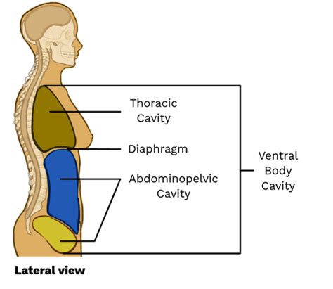
Table of Contents
- Name The Muscle That Subdivides The Ventral Body Cavity
- Table of Contents
- The Diaphragm: The Muscle That Subdivides the Ventral Body Cavity
- Understanding the Ventral Body Cavity
- 1. The Thoracic Cavity: A Sanctuary for Respiration and Circulation
- 2. The Abdominopelvic Cavity: The Digestive and Reproductive Hub
- The Diaphragm: The Master Divider
- Anatomy of the Diaphragm: A Detailed Look
- Function of the Diaphragm: Breathing and Beyond
- Clinical Significance of the Diaphragm: Conditions and Implications
- Conclusion: The Unsung Hero of the Ventral Body Cavity
- Latest Posts
- Latest Posts
- Related Post
The Diaphragm: The Muscle That Subdivides the Ventral Body Cavity
The human body is a marvel of intricate design, with various systems working in perfect harmony. One crucial aspect of this design is the organization of internal spaces, or body cavities. These cavities protect vital organs and allow for their efficient functioning. Among these, the ventral body cavity plays a significant role, housing many essential organs. Crucially, this large cavity is subdivided by a single, vital muscle: the diaphragm. This article will delve deep into the anatomy, function, and clinical significance of this remarkable muscle.
Understanding the Ventral Body Cavity
Before focusing on the diaphragm, let's establish a clear understanding of the ventral body cavity itself. The ventral body cavity, also known as the coelom, is a large, fluid-filled space that occupies the anterior aspect of the trunk. It's separated from the dorsal body cavity (containing the brain and spinal cord) by the vertebral column and associated muscles. The ventral body cavity's primary function is to protect the delicate organs it contains from external forces and to allow for their movement and expansion during processes like breathing and digestion.
The ventral body cavity is further subdivided into two major parts:
1. The Thoracic Cavity: A Sanctuary for Respiration and Circulation
Located superiorly, the thoracic cavity is encased by the rib cage, sternum, and associated muscles. It houses the heart, lungs, and major blood vessels, all crucial for respiration and circulation. The thoracic cavity is further partitioned into three smaller compartments:
- Mediastinum: The central compartment, separating the two lungs. It contains the heart, thymus gland, trachea, esophagus, and major blood vessels.
- Right Pleural Cavity: Surrounds the right lung.
- Left Pleural Cavity: Surrounds the left lung.
Each lung is enveloped by a double-layered serous membrane called the pleura. The visceral pleura clings tightly to the lung surface, while the parietal pleura lines the inner surface of the thoracic cavity. The pleural cavity, the space between the two layers, contains a small amount of serous fluid which minimizes friction during breathing.
2. The Abdominopelvic Cavity: The Digestive and Reproductive Hub
Inferior to the thoracic cavity, the abdominopelvic cavity is a larger space that houses the majority of the digestive, reproductive, and urinary organs. It's not completely separated from the thoracic cavity, but rather separated by the diaphragm. This cavity is further divided into two regions:
- Abdominal Cavity: The superior portion, containing the stomach, intestines, liver, spleen, pancreas, kidneys, and adrenal glands. It's primarily responsible for digestion and metabolism.
- Pelvic Cavity: The inferior portion, nestled within the bony pelvis. It contains the bladder, reproductive organs (uterus, ovaries, prostate gland), and the rectum.
The Diaphragm: The Master Divider
The diaphragm, a broad, dome-shaped muscle, acts as the crucial anatomical barrier separating the thoracic cavity from the abdominopelvic cavity. Its unique structure and function are essential for breathing, maintaining proper abdominal pressure, and supporting other physiological processes.
Anatomy of the Diaphragm: A Detailed Look
The diaphragm's anatomy is complex, yet elegantly designed for its vital function. Consider the following key aspects:
- Shape and Location: As mentioned, the diaphragm is a dome-shaped muscle. It's located at the base of the thoracic cavity, attaching to the inferior border of the rib cage, sternum, and lumbar vertebrae. Its central tendon is a strong, fibrous structure to which the muscle fibers converge.
- Muscle Fibers: The diaphragm’s muscle fibers originate from various points of attachment and converge on the central tendon. These fibers are arranged in a radial pattern, allowing for a coordinated contraction.
- Openings: Several important openings perforate the diaphragm, allowing structures like the esophagus, inferior vena cava, and aorta to pass between the thoracic and abdominopelvic cavities. These openings are strategically placed to prevent unwanted movement of organs or fluids.
- Innervation: The phrenic nerves, originating from the cervical spinal cord (C3-C5), provide motor innervation to the diaphragm. This is crucial for the diaphragm's voluntary and involuntary control.
Function of the Diaphragm: Breathing and Beyond
The diaphragm's primary role is in respiration. During inhalation, the diaphragm contracts, flattening its dome shape and increasing the volume of the thoracic cavity. This decrease in intrathoracic pressure draws air into the lungs. During exhalation, the diaphragm relaxes, returning to its dome shape and decreasing the thoracic cavity volume, forcing air out of the lungs. This process is largely involuntary, regulated by the respiratory centers in the brainstem. However, the diaphragm also exhibits voluntary control, allowing for activities like singing and forceful exhalation (coughing).
Beyond respiration, the diaphragm plays a significant role in:
- Abdominal Pressure Regulation: The diaphragm's contraction assists in increasing intra-abdominal pressure. This is crucial for functions such as defecation, urination, and childbirth.
- Venous Return: The diaphragm's movement aids in venous return by compressing abdominal veins, pushing blood back towards the heart.
- Coughing and Sneezing: The forceful contractions of the diaphragm are vital for generating the pressure needed for coughing and sneezing.
- Hiccups: A sudden, involuntary contraction of the diaphragm causes the characteristic hiccup sound.
Clinical Significance of the Diaphragm: Conditions and Implications
The diaphragm's importance is highlighted by the various clinical conditions that can affect its function. Consider these:
- Diaphragmatic Hernia: A hole in the diaphragm allows abdominal organs to move into the thoracic cavity. This can cause respiratory distress and other complications, often requiring surgical repair.
- Diaphragmatic Paralysis: Damage to the phrenic nerves can result in paralysis of the diaphragm, significantly impacting breathing and requiring respiratory support. This can be caused by various factors such as trauma, surgery, or diseases.
- Diaphragmatic Eventration: A weakening of the diaphragm leads to its upward displacement. This can cause shortness of breath and compromise respiratory function.
- Hiatal Hernia: A condition where a portion of the stomach protrudes through the esophageal hiatus (opening in the diaphragm for the esophagus). This can lead to heartburn, acid reflux, and other digestive problems.
- Respiratory Infections: Because the diaphragm is so close to other organs, infections can impact its function.
Conclusion: The Unsung Hero of the Ventral Body Cavity
The diaphragm, often overlooked, plays a crucial role in the human body. As the primary muscle dividing the thoracic and abdominopelvic cavities, it’s essential for respiration, regulating intra-abdominal pressure, and facilitating various other vital bodily functions. Understanding its anatomy and function is essential for appreciating the intricate workings of the human body and for diagnosing and treating conditions affecting this vital muscle. Its complex structure and multifunctional role emphasize its importance as a critical component of our overall health and well-being. Further research into the diaphragm's role in various physiological processes continues to unveil its multifaceted contributions to our body’s homeostasis and overall efficiency. This remarkable muscle, indeed, deserves recognition as the unsung hero of the ventral body cavity.
Latest Posts
Latest Posts
-
Which Of The Following Is Not A Steroid Hormone
Mar 24, 2025
-
The Layer Of Gases Surrounding Earth Is The
Mar 24, 2025
-
What Is 3 Percent Of 18
Mar 24, 2025
-
In Triangle Abc The Measure Of Angle B Is 90
Mar 24, 2025
-
Write The Iupac Name Of The Compound Shown
Mar 24, 2025
Related Post
Thank you for visiting our website which covers about Name The Muscle That Subdivides The Ventral Body Cavity . We hope the information provided has been useful to you. Feel free to contact us if you have any questions or need further assistance. See you next time and don't miss to bookmark.
