Microscopic Study Of Tissues Is Called
News Leon
Mar 29, 2025 · 7 min read
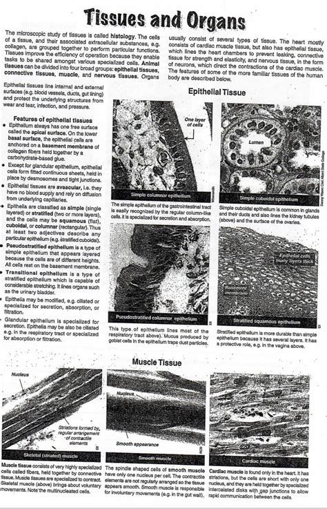
Table of Contents
Microscopic Study of Tissues: A Deep Dive into Histology
The microscopic study of tissues is called histology. This fascinating field provides a crucial bridge between the macroscopic anatomy of the body and the intricate molecular processes that govern its function. Histologists utilize a variety of techniques to prepare, stain, and examine tissue samples, revealing the cellular architecture and providing insights into both health and disease. This comprehensive article will delve into the intricacies of histology, exploring its techniques, applications, and significance in medical diagnostics and research.
What is Histology?
Histology, derived from the Greek words histos (tissue) and logos (study), is the branch of biology that deals with the microscopic structure of tissues. It’s a cornerstone of many medical and biological disciplines, offering a window into the microscopic world of cells and their organization into functional units. Understanding histology is critical for comprehending the normal structure of tissues and organs, and equally vital for diagnosing disease. Pathologists, for instance, rely heavily on histological analysis to identify cancerous growths, infections, and other abnormalities.
The Importance of Tissue Preparation
Before a tissue sample can be examined under a microscope, it must undergo a careful preparation process. This process, which is crucial for obtaining high-quality images and accurate results, typically involves several key steps:
1. Fixation: Preserving Tissue Integrity
Fixation is the initial critical step, aiming to preserve the tissue's structure and prevent degradation. Common fixatives include formalin (formaldehyde), which cross-links proteins, and alcohol, which dehydrates the tissue. The choice of fixative depends on the specific tissue type and the intended staining methods. Improper fixation can lead to artifacts – distortions or abnormalities in the tissue – that can compromise the accuracy of the histological analysis.
2. Processing: Embedding the Tissue
Following fixation, the tissue undergoes processing to prepare it for sectioning. This involves a series of steps, typically involving dehydration using graded alcohols, clearing with solvents like xylene, and infiltration with paraffin wax. Paraffin wax provides a firm support medium for the tissue, enabling the creation of thin sections suitable for microscopic examination.
3. Sectioning: Creating Thin Tissue Slices
The paraffin-embedded tissue block is then sectioned using a microtome, a specialized instrument that creates extremely thin slices (typically 3-5 micrometers thick). These thin sections are then mounted onto glass slides, ready for staining. The thickness of the section is crucial; too thick, and details will be obscured; too thin, and the section may be fragile.
4. Staining: Enhancing Visibility
The tissue sections are colorless when mounted on slides and require staining to enhance the visibility of cellular structures and components. Various stains are available, each with its own specific affinity for certain cellular components. Hematoxylin and eosin (H&E) staining is the most common technique, with hematoxylin staining the cell nuclei blue/purple and eosin staining the cytoplasm pink/red. Other specialized stains, such as periodic acid-Schiff (PAS) for carbohydrates and Masson's trichrome for connective tissue, reveal specific structures and components. The choice of stain is guided by the specific histological question being addressed.
Types of Tissues Studied in Histology
Histology encompasses the study of four primary tissue types:
1. Epithelial Tissue: Covering and Lining
Epithelial tissues are sheets of tightly packed cells that cover body surfaces, line body cavities, and form glands. They are classified based on cell shape (squamous, cuboidal, columnar) and layering (simple, stratified, pseudostratified). Histology reveals the distinct arrangements of cells in different epithelial tissues, reflecting their specialized functions, such as protection, secretion, absorption, and excretion. For example, the stratified squamous epithelium of the skin provides a tough, protective barrier, while the simple columnar epithelium of the small intestine is specialized for absorption.
2. Connective Tissue: Support and Connection
Connective tissues are diverse in structure and function, providing support, connecting tissues, and transporting substances. They are characterized by a relatively abundant extracellular matrix, containing various proteins like collagen and elastin, along with ground substance. Histology reveals the arrangement of cells and the composition of the extracellular matrix in different connective tissues, such as loose connective tissue, dense regular connective tissue (tendons, ligaments), adipose tissue, cartilage, and bone. The variations in matrix composition reflect the differing mechanical properties of these tissues.
3. Muscle Tissue: Movement and Contraction
Muscle tissues are responsible for movement and are characterized by specialized contractile cells. Histology distinguishes three types of muscle tissue: skeletal muscle (voluntary, striated), smooth muscle (involuntary, non-striated), and cardiac muscle (involuntary, striated). The microscopic appearance of each type of muscle tissue reflects its unique functional properties. Skeletal muscle shows prominent striations due to the organized arrangement of actin and myosin filaments, whereas smooth muscle lacks striations. Cardiac muscle has intercalated discs, specialized junctions that facilitate rapid communication between cardiac muscle cells.
4. Nervous Tissue: Communication and Control
Nervous tissue is specialized for communication and control, comprising neurons (nerve cells) and supporting glial cells. Histology reveals the characteristic morphology of neurons, including their cell bodies, dendrites, and axons. The organization of neurons and glial cells into complex networks within the brain and spinal cord is crucial for information processing and transmission. Studying the structure of synapses, the junctions between neurons, is vital for understanding neural communication.
Techniques Used in Histology
Beyond basic staining, several advanced techniques enhance the visualization and analysis of tissues:
1. Immunohistochemistry (IHC): Identifying Specific Proteins
Immunohistochemistry utilizes antibodies to detect specific proteins within tissues. Antibodies, which are highly specific to their target proteins, are labeled with enzymes or fluorescent molecules, allowing visualization of the target protein's location within the tissue. IHC is invaluable in diagnosing diseases such as cancer, where the expression of specific proteins can indicate the type and grade of the tumor.
2. In Situ Hybridization (ISH): Detecting Specific Nucleic Acids
In situ hybridization is a technique used to detect specific nucleic acid sequences (DNA or RNA) within tissues. Labeled probes, complementary to the target sequence, bind to the target nucleic acid, enabling visualization of its location. ISH is useful for detecting viral or bacterial infections, genetic mutations, and gene expression patterns.
3. Electron Microscopy: High-Resolution Imaging
Electron microscopy offers much higher resolution than light microscopy, enabling visualization of subcellular structures. Transmission electron microscopy (TEM) provides cross-sectional images of cells, revealing internal organelles in detail. Scanning electron microscopy (SEM) provides three-dimensional images of the surfaces of tissues and cells.
4. Confocal Microscopy: 3D Imaging of Tissues
Confocal microscopy uses lasers and optical sectioning to create high-resolution images of tissues, allowing for the reconstruction of three-dimensional structures. This technique is especially useful for studying complex tissues such as the nervous system, where neurons form intricate networks.
Applications of Histology
Histology's applications span numerous fields:
1. Medical Diagnosis: Identifying Diseases
Histological examination of biopsy samples is crucial for diagnosing a wide range of diseases, including cancer, infections, and inflammatory conditions. Pathologists analyze tissue samples to identify abnormal cells, tissue architecture, and inflammatory responses, providing vital information for diagnosis and treatment planning. The accurate interpretation of histological findings is paramount in guiding clinical decisions.
2. Research: Understanding Biological Processes
Histology plays a vital role in biological research, providing insights into the structure and function of tissues and organs in both health and disease. Researchers use histological techniques to investigate the effects of drugs, toxins, and environmental factors on tissues, and to study developmental processes and cellular interactions.
3. Forensic Science: Investigating Crime Scenes
Histology can be used in forensic investigations to analyze tissue samples, providing information relevant to identifying individuals, determining the cause of death, and reconstructing events.
4. Veterinary Medicine: Diagnosing Animal Diseases
Histological techniques are also applied in veterinary medicine for diagnosing diseases in animals. Histological examination of tissue samples from animals can help identify infections, tumors, and other abnormalities.
The Future of Histology
Technological advancements are continuously shaping the future of histology. Digital pathology, which involves the use of digital images of tissue sections, is becoming increasingly prevalent, allowing for remote access to samples and facilitating collaborative diagnosis and research. Advances in microscopy techniques, such as super-resolution microscopy, are pushing the boundaries of resolution, enabling even greater detail in the visualization of cellular structures. The integration of artificial intelligence and machine learning is also transforming histological analysis, providing automated tools for image analysis and diagnosis.
In conclusion, the microscopic study of tissues, or histology, is a fundamental discipline with far-reaching applications across medicine, biology, and other fields. Its techniques provide invaluable insights into the structure and function of tissues, crucial for understanding health and diagnosing disease. As technology continues to advance, histology's role in advancing our understanding of the biological world will undoubtedly continue to grow.
Latest Posts
Latest Posts
-
Which Organelle Is Enclosed By A Double Membrane
Mar 31, 2025
-
Compare And Contrast An Ecosystem And A Habitat
Mar 31, 2025
-
Network Layer Firewall Works As A
Mar 31, 2025
-
Is Sodium Methoxide A Strong Nucleophile
Mar 31, 2025
-
The Amount Of Space An Object Occupies
Mar 31, 2025
Related Post
Thank you for visiting our website which covers about Microscopic Study Of Tissues Is Called . We hope the information provided has been useful to you. Feel free to contact us if you have any questions or need further assistance. See you next time and don't miss to bookmark.
