Label The Parts Of The Nephron
News Leon
Mar 31, 2025 · 6 min read
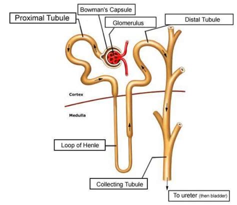
Table of Contents
Label the Parts of the Nephron: A Comprehensive Guide
The nephron, the functional unit of the kidney, is a complex structure responsible for filtering blood and producing urine. Understanding its intricate parts is crucial to grasping the intricacies of renal physiology. This comprehensive guide will delve into the detailed anatomy of the nephron, meticulously labeling each component and explaining its role in the urinary system. We'll explore the process of filtration, reabsorption, and secretion, highlighting the specific contributions of each nephron segment.
The Two Main Parts of the Nephron: A Bird's Eye View
Before we dive into the specifics, it's essential to understand the nephron's fundamental structure. The nephron is broadly divided into two main parts:
- Renal Corpuscle: This is the initial filtering unit, composed of the glomerulus and Bowman's capsule.
- Renal Tubule: This long, convoluted tube processes the filtrate from the renal corpuscle, modifying its composition before it becomes urine.
Detailed Anatomy and Function of the Renal Corpuscle
The renal corpuscle, the site of blood filtration, is comprised of:
1. Glomerulus: The Filtration Bed
The glomerulus is a network of capillaries nestled within Bowman's capsule. Its unique structure facilitates efficient filtration:
- Fenestrated Endothelial Cells: The capillary walls are perforated by numerous pores (fenestrations), allowing water and small solutes to pass through while restricting larger proteins and blood cells.
- Glomerular Basement Membrane (GBM): This specialized layer acts as a selective filter, preventing the passage of larger molecules and negatively charged proteins. Its composition of collagen, laminin, and proteoglycans contributes to its selective permeability.
- Podocytes: These specialized epithelial cells surround the glomerular capillaries. Their intricate foot processes (pedicels) interdigitate, forming filtration slits further refining the filtration process. These slits are covered by a thin diaphragm, further regulating what passes through.
The high hydrostatic pressure within the glomerular capillaries drives the filtration process, forcing fluid and solutes into Bowman's capsule.
2. Bowman's Capsule: The Collecting Chamber
Bowman's capsule is a double-walled cup-like structure surrounding the glomerulus. It receives the filtrate produced by the glomerulus and directs it into the renal tubule.
- Parietal Layer: The outer layer of Bowman's capsule is a simple squamous epithelium, providing structural support.
- Visceral Layer: The inner layer is composed of the podocytes mentioned above, actively participating in the filtration process.
- Bowman's Space: The space between the parietal and visceral layers, where the filtrate collects before entering the renal tubule.
Detailed Anatomy and Function of the Renal Tubule
The renal tubule is a long, winding tube responsible for modifying the filtrate through reabsorption and secretion. It is divided into several distinct segments:
1. Proximal Convoluted Tubule (PCT): Reabsorption Central
The PCT is the initial segment of the renal tubule, characterized by its highly convoluted structure. It plays a vital role in reabsorption:
- Extensive Brush Border: The apical surface of the PCT cells possesses numerous microvilli, forming a brush border that significantly increases the surface area for reabsorption.
- Abundant Mitochondria: The high metabolic activity of the PCT requires abundant mitochondria to fuel the active transport processes involved in reabsorption.
- Reabsorption of Essential Substances: The PCT reabsorbs the majority of essential nutrients (glucose, amino acids), electrolytes (sodium, potassium, chloride), and water. This reabsorption is driven by both active and passive transport mechanisms.
The PCT also secretes certain substances like hydrogen ions (H+) and ammonia (NH3) into the tubular fluid.
2. Loop of Henle: Establishing the Concentration Gradient
The Loop of Henle extends from the PCT into the renal medulla and back to the cortex. It plays a crucial role in establishing the medullary osmotic gradient, which is essential for concentrating urine:
- Descending Limb: This limb is permeable to water but relatively impermeable to solutes. Water passively moves out of the descending limb, driven by the high osmolarity of the medullary interstitium.
- Ascending Limb: This limb is impermeable to water but actively transports sodium, potassium, and chloride ions out of the tubule. This active transport contributes to the hyperosmolarity of the medullary interstitium.
The countercurrent multiplier system, involving the interaction between the descending and ascending limbs, amplifies the osmotic gradient, allowing the kidneys to produce highly concentrated urine.
3. Distal Convoluted Tubule (DCT): Fine-Tuning the Filtrate
The DCT is the final segment of the renal tubule before the collecting duct. It plays a crucial role in fine-tuning the composition of the filtrate:
- Sodium Reabsorption: Sodium reabsorption in the DCT is regulated by aldosterone, a hormone produced by the adrenal cortex. Aldosterone stimulates sodium reabsorption, increasing water reabsorption and blood pressure.
- Potassium Secretion: Potassium secretion in the DCT is also regulated by aldosterone. Aldosterone stimulates potassium secretion, maintaining potassium homeostasis.
- Calcium Reabsorption: Parathyroid hormone (PTH) stimulates calcium reabsorption in the DCT, regulating calcium balance in the body.
4. Collecting Duct: Concentrating Urine
The collecting duct receives the filtrate from several nephrons and plays a critical role in regulating urine concentration and excretion:
- Water Reabsorption: The permeability of the collecting duct to water is regulated by antidiuretic hormone (ADH), also known as vasopressin. ADH increases water permeability, allowing water to be reabsorbed and producing concentrated urine.
- Acid-Base Balance: The collecting duct contributes to acid-base balance by secreting hydrogen ions (H+) and reabsorbing bicarbonate ions (HCO3-).
Types of Nephrons: Cortical and Juxtamedullary
Nephrons are further classified based on their location and the length of their Loop of Henle:
- Cortical Nephrons: These nephrons are located primarily in the cortex and have short Loops of Henle that extend only slightly into the medulla. They are responsible for most of the filtration and reabsorption of nutrients and electrolytes.
- Juxtamedullary Nephrons: These nephrons have long Loops of Henle that extend deep into the medulla. They play a crucial role in concentrating urine and maintaining water balance. Their longer loops contribute significantly to the countercurrent multiplier system.
The Juxtaglomerular Apparatus (JGA): Regulation of Blood Pressure
The JGA is a specialized structure located where the afferent arteriole and the distal convoluted tubule come into contact. It plays a crucial role in regulating blood pressure:
- Juxtaglomerular Cells: Modified smooth muscle cells in the afferent arteriole that synthesize and release renin. Renin is an enzyme that initiates the renin-angiotensin-aldosterone system (RAAS), which helps to regulate blood pressure.
- Macula Densa: Specialized cells in the distal convoluted tubule that detect changes in sodium concentration in the tubular fluid. They signal juxtaglomerular cells to release renin if sodium concentration is low.
Clinical Significance: Understanding Nephron Dysfunction
Understanding the structure and function of the nephron is crucial for diagnosing and treating various kidney diseases. Damage to the nephrons can lead to impaired filtration, reabsorption, and secretion, resulting in conditions like:
- Glomerulonephritis: Inflammation of the glomeruli, leading to proteinuria (protein in urine) and hematuria (blood in urine).
- Acute Kidney Injury (AKI): Sudden loss of kidney function, often caused by infection, dehydration, or medications.
- Chronic Kidney Disease (CKD): Progressive loss of kidney function over time, often caused by diabetes, hypertension, or glomerulonephritis.
Conclusion: The Intricate Machinery of Filtration and Excretion
The nephron's intricate structure reflects its multifaceted role in maintaining homeostasis. By understanding the functions of each component – from the glomerulus to the collecting duct – we gain a deeper appreciation for the complex processes involved in blood filtration, reabsorption, secretion, and urine concentration. This detailed knowledge is not only essential for basic biological understanding but also forms the cornerstone of diagnosing and managing various renal disorders. The study of the nephron underscores the remarkable efficiency and precision of the human body's regulatory mechanisms. Further exploration into specific transporter proteins, hormonal influences, and the intricate interplay between different nephron segments will only deepen our comprehension of this vital organ's operation.
Latest Posts
Latest Posts
-
Experiment To Show That Chlorophyll Is Necessary For Photosynthesis Meritnation
Apr 02, 2025
-
What Is The Difference Between Heterochromatin And Euchromatin
Apr 02, 2025
-
In Order List The Steps Of The Scientific Method
Apr 02, 2025
-
How To Find Instantaneous Rate Of Change From A Table
Apr 02, 2025
-
Which Of The Following Will Have The Highest Boiling Point
Apr 02, 2025
Related Post
Thank you for visiting our website which covers about Label The Parts Of The Nephron . We hope the information provided has been useful to you. Feel free to contact us if you have any questions or need further assistance. See you next time and don't miss to bookmark.
