Inspiratory And Expiratory Centers Are Located In The
News Leon
Mar 17, 2025 · 6 min read
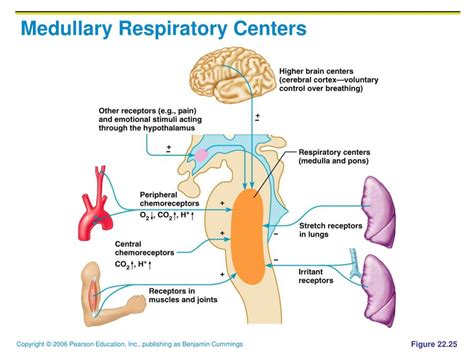
Table of Contents
Inspiratory and Expiratory Centers are Located in the Medulla Oblongata and Pons: A Deep Dive into Respiratory Control
The rhythmic, seemingly effortless act of breathing is far from simple. Behind each inhale and exhale lies a complex interplay of neural circuits, chemical messengers, and mechanical processes meticulously orchestrated to maintain life. Understanding the control of respiration requires understanding the location and function of the inspiratory and expiratory centers. This article will delve deep into the neural structures responsible for our breath, exploring their location within the brainstem, their intricate interactions, and the factors influencing their activity.
The Brainstem Respiratory Centers: A Neurological Orchestra
The primary control centers for breathing reside within the brainstem, specifically the medulla oblongata and the pons. These aren't isolated entities; rather, they represent a network of interconnected neurons working in concert. Let's examine the key components:
1. Medullary Respiratory Centers: The Primary Conductors
The medulla oblongata houses the two crucial centers responsible for the basic rhythm of breathing:
-
Dorsal Respiratory Group (DRG): This group of neurons, located within the dorsal medulla, plays a pivotal role in inspiration. It receives sensory input from various peripheral chemoreceptors and mechanoreceptors (more on this later) and sends signals to the diaphragm via the phrenic nerve and to the intercostal muscles via intercostal nerves, initiating inspiration. The DRG’s activity is primarily responsible for the basic rhythm of breathing, setting the pace for the respiratory cycle. Think of the DRG as the metronome of breathing.
-
Ventral Respiratory Group (VRG): Situated in the ventral medulla, the VRG is more complex and multifaceted than the DRG. While largely inactive during quiet breathing, it becomes crucial during forceful breathing (e.g., exercise, gasping). The VRG contains both inspiratory and expiratory neurons. Its inspiratory neurons can augment the activity of the DRG during increased respiratory demand. The expiratory neurons in the VRG activate the expiratory muscles, like the abdominal muscles and internal intercostals, during forceful exhalation. The VRG acts as the amplifier of the respiratory system, kicking into high gear when needed.
2. Pontine Respiratory Centers: The Fine-Tuners
The pons, located superior to the medulla, houses respiratory centers that refine and modulate the basic rhythm generated by the medullary centers. These centers don't initiate breathing but influence the timing and depth of breaths:
-
Pneumotaxic Center: Located in the upper pons, this center limits inspiration, preventing overinflation of the lungs. It acts as a "switch," shortening the duration of inspiration and influencing the respiratory rate. Think of it as the conductor subtly adjusting the tempo of the orchestra.
-
Apneustic Center: Located in the lower pons, this center prolongs inspiration. It counteracts the pneumotaxic center, ensuring that inspiration continues for a sufficient duration. A balance between these two pontine centers is crucial for regulating the depth and frequency of breathing. The apneustic center can be seen as the counterbalance to the pneumotaxic center, ensuring a balanced and effective respiratory cycle.
Sensory Input: The Feedback Loop
The respiratory centers don't operate in isolation; they constantly receive feedback from various sensory receptors throughout the body, ensuring accurate and responsive regulation of breathing.
1. Peripheral Chemoreceptors: Monitoring Blood Chemistry
These receptors, located in the carotid bodies (at the bifurcation of the common carotid arteries) and aortic bodies (in the aortic arch), are exquisitely sensitive to changes in blood oxygen (O2), carbon dioxide (CO2), and pH.
- Decreased O2 (hypoxemia): Stimulates increased ventilation to boost oxygen uptake.
- Increased CO2 (hypercapnia): The most potent stimulus for increased ventilation. CO2 dissolves in blood, forming carbonic acid, lowering pH.
- Decreased pH (acidosis): Stimulates increased ventilation to expel CO2 and raise pH.
These chemoreceptors constantly monitor blood composition and send signals to the medullary respiratory centers, fine-tuning breathing to maintain optimal blood gas levels.
2. Central Chemoreceptors: Monitoring Brain Cerebrospinal Fluid (CSF)
Located in the medulla, these chemoreceptors are sensitive to changes in the partial pressure of CO2 (PCO2) in the cerebrospinal fluid (CSF). CO2 readily diffuses from the blood into the CSF, where it affects pH. Increased PCO2 in the CSF leads to decreased pH, stimulating central chemoreceptors and consequently increasing ventilation.
3. Mechanoreceptors: Monitoring Lung Stretch and Airflow
These receptors provide feedback about the mechanical state of the lungs and airways:
- Stretch Receptors (pulmonary stretch receptors): Located in the airways and lung parenchyma, these receptors are sensitive to lung inflation. When the lungs are overinflated, they send signals to the pneumotaxic center, inhibiting further inspiration (Hering-Breuer reflex). This prevents overstretching and damage to the lungs.
- Irritant Receptors: Located in the airways, these receptors are sensitive to irritants like dust, smoke, or excess mucus. They trigger a cough or bronchoconstriction to remove the irritant.
- J-receptors (juxtacapillary receptors): Located in the interstitial tissue of the lungs, these receptors respond to alveolar congestion and edema. They can trigger rapid, shallow breathing.
Higher Brain Centers: Voluntary Control and Emotional Influence
While the brainstem centers control the automatic rhythm of breathing, higher brain centers can exert voluntary control over respiration. The cerebral cortex allows conscious control of breathing, such as holding your breath or taking deep breaths. However, this voluntary control is limited; overriding the brainstem centers for prolonged periods can be dangerous.
Furthermore, emotions also influence breathing. For instance, stress, fear, or anxiety can lead to rapid, shallow breathing (hyperventilation). Conversely, relaxation can lead to slower, deeper breaths. These emotional influences involve connections between the limbic system (involved in emotions) and the brainstem respiratory centers.
Clinical Significance: Respiratory Disorders
Understanding the location and function of the respiratory centers is crucial for diagnosing and treating various respiratory disorders. Dysfunction in these centers or their connections can lead to:
- Apnea: Cessation of breathing. Central apnea originates from dysfunction within the brainstem respiratory centers.
- Hyperventilation: Rapid, deep breathing, often due to anxiety or metabolic acidosis.
- Hypoventilation: Slow, shallow breathing, leading to increased CO2 levels.
- Cheyne-Stokes Respiration: Alternating periods of apnea and hyperventilation, often associated with heart failure or brain injury.
- Respiratory Distress Syndrome: Characterized by impaired gas exchange, often affecting premature infants due to underdeveloped lungs.
Conclusion: A Breathtakingly Complex System
The seemingly simple act of breathing is orchestrated by a remarkably complex interplay of neural structures and feedback mechanisms. The inspiratory and expiratory centers, located in the medulla oblongata and pons, work in concert with sensory receptors and higher brain centers to regulate the rhythm, depth, and pattern of breathing. A thorough understanding of these centers is essential for comprehending the physiological basis of respiration and various related clinical conditions. Further research into the intricate workings of this system is crucial for developing better diagnostics and treatments for respiratory diseases and disorders. The ongoing exploration of the respiratory system's intricacies unveils a continuous evolution of our understanding of this fundamental aspect of human life. From the basic rhythmic control to the complex interplay of feedback mechanisms and higher-level influence, the journey of understanding respiration is far from over, promising further discoveries and breakthroughs in the years to come.
Latest Posts
Latest Posts
-
How Many Oxygen Molecules Can One Hemoglobin Carry
Mar 18, 2025
-
Which Of The Following Is Not A Form Of Precipitation
Mar 18, 2025
-
Which Statement About Natural Selection Is True
Mar 18, 2025
-
Which Chamber Of Heart Has Thickest Wall
Mar 18, 2025
-
How Many Feet Is 1 2 Miles
Mar 18, 2025
Related Post
Thank you for visiting our website which covers about Inspiratory And Expiratory Centers Are Located In The . We hope the information provided has been useful to you. Feel free to contact us if you have any questions or need further assistance. See you next time and don't miss to bookmark.
