Fuse To Form The Coxal Bone
News Leon
Mar 18, 2025 · 8 min read
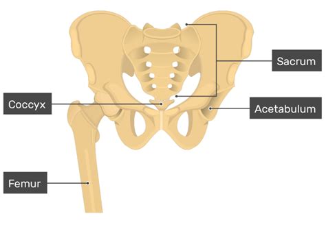
Table of Contents
The Fusion of Bones: Forming the Coxal Bone (Hip Bone)
The coxal bone, also known as the hip bone or innominate bone, is a large, complex structure that plays a crucial role in weight-bearing, locomotion, and protecting internal organs. Unlike many other bones that develop from a single ossification center, the coxal bone is formed through the intricate fusion of three separate bones: the ilium, ischium, and pubis. This fusion process is a fascinating example of developmental biology, and understanding its intricacies is key to appreciating the strength and functionality of the adult hip.
The Three Primary Bones: Ilium, Ischium, and Pubis
Before delving into the fusion process itself, let's examine the three individual bones that contribute to the coxal bone:
1. The Ilium: The Superior Bone
The ilium is the largest of the three bones and forms the superior portion of the coxal bone. Its wing-like structure, the ala, is broad and flared, providing a substantial surface area for muscle attachment. Key features of the ilium include:
- Iliac Crest: The superior border of the ilium, easily palpable on the surface of the body. This serves as an important landmark for anatomical reference.
- Iliac Fossa: A concave area on the internal surface of the ilium, providing attachment points for several hip muscles.
- Auricular Surface: A roughened area on the posterior aspect of the ilium, which articulates with the sacrum to form the sacroiliac joint.
- Greater Sciatic Notch: A large notch located on the posterior border of the ilium, which contributes to the formation of the sciatic foramen.
2. The Ischium: The Inferior and Posterior Bone
The ischium forms the inferior and posterior portion of the coxal bone. It's characterized by its strong, robust construction, reflecting its role in supporting body weight during sitting. Important features include:
- Ischial Tuberosity: The roughened, weight-bearing area at the inferior end of the ischium. This is the part of the bone that comes into contact with a surface when sitting.
- Ischial Spine: A prominent projection located superior to the ischial tuberosity, serving as a landmark and attachment point for ligaments and muscles.
- Lesser Sciatic Notch: A smaller notch located superior to the ischial tuberosity, contributing to the sciatic foramen.
3. The Pubis: The Anterior Bone
The pubis forms the anterior portion of the coxal bone. It contributes to the formation of the pubic symphysis, the joint that connects the two coxal bones at the anterior midline of the pelvis. Key anatomical features of the pubis are:
- Superior Ramus: The superior branch of the pubis, extending towards the ilium.
- Inferior Ramus: The inferior branch of the pubis, extending towards the ischium.
- Pubic Crest: The superior border of the pubic body, which forms part of the pubic symphysis.
- Pubic Tubercle: A bony projection located at the junction of the superior and inferior rami, an important landmark and muscle attachment point.
The Fusion Process: From Three to One
The fusion of the ilium, ischium, and pubis into a single coxal bone is a complex process that begins during fetal development and continues into adolescence. The process is primarily driven by endochondral ossification, which involves the gradual replacement of cartilage with bone tissue.
Stages of Fusion:
-
Early Development: In the early stages of fetal development, the ilium, ischium, and pubis exist as separate cartilaginous structures. These cartilaginous elements gradually undergo ossification, with primary ossification centers appearing within each bone.
-
Secondary Ossification Centers: As the bones grow and develop, secondary ossification centers appear at various locations within each bone. These centers contribute to the overall growth and shape of the individual bones.
-
Triradiate Cartilage: The three bones are initially connected by strips of cartilage known as the triradiate cartilage. This cartilage persists throughout childhood and adolescence, providing a growth plate that allows for continued bone growth.
-
Fusion Begins: During puberty and adolescence, the triradiate cartilage begins to gradually ossify and fuse. This fusion process starts at the points of contact between the three bones and progresses outwards, eventually resulting in a single, unified coxal bone.
-
Complete Fusion: Complete fusion of the three bones typically occurs around the age of 15-17 years in females and 17-19 years in males. However, this can vary, and slight variations in the fusion timeline are considered normal.
The Acetabulum: The Hip Socket
A crucial aspect of the coxal bone's formation is the development of the acetabulum, the deep, cup-shaped socket that articulates with the head of the femur to form the hip joint. The acetabulum is formed by the contributions of all three constituent bones: the ilium, ischium, and pubis. Its smooth, articular surface provides a stable and efficient joint for weight-bearing and movement. The depth of the acetabulum, along with the surrounding ligaments and muscles, contributes to the stability and strength of the hip joint.
Clinical Significance: Implications of Incomplete Fusion
Incomplete fusion of the coxal bone, although relatively rare, can lead to several clinical issues. These issues can range from subtle discomfort and limited mobility to more significant problems requiring surgical intervention. Some of the potential implications include:
- Osteoarthritis: An incomplete fusion may disrupt the normal biomechanics of the hip joint, increasing the risk of early onset osteoarthritis.
- Hip Instability: The weakened structure from incomplete fusion can lead to hip instability and an increased risk of dislocation.
- Pain and Discomfort: Persistent pain and discomfort in the hip region can be a result of incomplete fusion or related biomechanical issues.
- Fractures: The area of incomplete fusion may be more prone to fractures compared to a fully fused bone.
Diagnosis of incomplete fusion typically involves imaging techniques like X-rays, which can visualize the extent of fusion or the presence of persistent cartilage. Treatment options depend on the severity of the condition, ranging from conservative measures like physical therapy to surgical intervention in more severe cases.
The Coxal Bone: A Strong and Adaptable Structure
The fusion of the ilium, ischium, and pubis to form the coxal bone is a testament to the complexity and efficiency of biological processes. This intricate developmental process results in a strong, adaptable structure that plays a critical role in supporting our bodies and enabling locomotion. Understanding the stages of fusion, the contributions of each constituent bone, and the potential clinical implications of incomplete fusion provides valuable insight into the human musculoskeletal system. Further research continues to illuminate the finer details of this remarkable developmental event, contributing to improved understanding of hip health and treatment.
Muscle Attachments and Functional Significance:
The coxal bone serves as a crucial attachment point for numerous muscles, making it pivotal for a wide range of movements including:
-
Hip flexion, extension, abduction, adduction, internal and external rotation: These fundamental movements are facilitated by muscles originating from or inserting onto the coxal bone, such as the gluteus maximus, gluteus medius, gluteus minimus, iliacus, psoas major, adductor muscles, and many more.
-
Trunk stabilization: The powerful muscles attached to the coxal bone play a vital role in stabilizing the trunk and maintaining posture. The deep abdominal muscles and the muscles of the back work in concert with the hip muscles for optimal spinal stability.
-
Locomotion: The coxal bone, along with the femur and surrounding musculature, forms the foundation of bipedal locomotion. The efficiency of weight transfer and limb movement depends on the integrity and functionality of the coxal bone.
-
Weight-bearing: The coxal bone is essential for weight-bearing, transmitting the body's weight from the upper body to the lower limbs during standing, walking, and running. The strong structure of the bone, together with the reinforcing ligaments and cartilage, ensures that it can withstand the considerable stresses involved in these activities.
The intricate arrangement of muscle attachments on the coxal bone, combined with the robust bone structure itself, makes it a remarkable example of biological engineering. The ability of the hip to efficiently execute a wide variety of movements and bear substantial loads is critical to the overall functionality of the human body.
Variations and Individual Differences:
While the general process of coxal bone fusion is consistent across individuals, subtle variations in timing and final morphology are common. Factors like genetics, nutrition, and overall health can influence the rate of bone growth and fusion. These individual differences are generally within a normal range and do not usually cause significant clinical issues. However, significant deviations from the typical pattern may warrant further investigation.
Furthermore, subtle asymmetries between the right and left coxal bones are frequently observed. These asymmetries are often related to factors such as handedness, habitual posture, and physical activities. They generally do not indicate any underlying pathology, but they demonstrate the plasticity of the musculoskeletal system in response to individual lifestyles and experiences.
Further Research and Future Directions:
Continued research into the intricacies of coxal bone fusion and its potential clinical implications remains crucial. Studies focusing on the molecular mechanisms regulating cartilage ossification, the impact of genetic factors, and the long-term consequences of variations in fusion timing are all active areas of investigation. A deeper understanding of these factors will lead to more precise diagnosis and tailored treatment approaches for individuals experiencing problems related to hip development or function. Advances in imaging technologies and biomechanical modeling are also contributing to a more comprehensive understanding of this complex structure and its role in human health. The continuous quest for knowledge in this area will undoubtedly lead to advancements in the prevention, diagnosis, and treatment of hip-related disorders.
Latest Posts
Latest Posts
-
Two Same Words With Different Meanings
Mar 18, 2025
-
Select The Correct Statement About Equilibrium
Mar 18, 2025
-
Draw The Major Product Of The Following Reaction
Mar 18, 2025
-
A Wire Loop Of Radius 10 Cm And Resistance
Mar 18, 2025
-
How Many Water Molecules In A Drop Of Water
Mar 18, 2025
Related Post
Thank you for visiting our website which covers about Fuse To Form The Coxal Bone . We hope the information provided has been useful to you. Feel free to contact us if you have any questions or need further assistance. See you next time and don't miss to bookmark.
