Correctly Identify The Following Formed Elements.
News Leon
Mar 19, 2025 · 6 min read
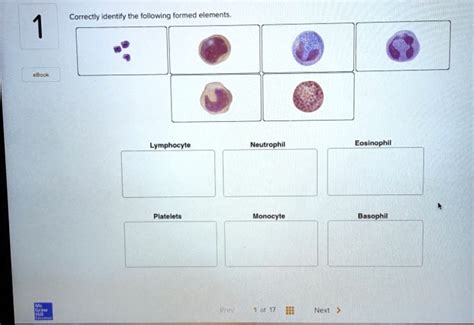
Table of Contents
Correctly Identify the Following Formed Elements: A Comprehensive Guide
Identifying formed elements in blood is a crucial skill in hematology and related fields. These elements, also known as blood cells, are responsible for a wide array of vital functions, from oxygen transport and immune defense to blood clotting. Accurate identification requires a keen understanding of their morphology, staining characteristics, and relative abundance. This comprehensive guide will delve into the correct identification of the major formed elements, providing detailed descriptions and high-yield information for students and professionals alike.
Erythrocytes (Red Blood Cells)
Erythrocytes, or red blood cells (RBCs), are the most numerous formed elements in blood. Their primary function is the transport of oxygen from the lungs to the body's tissues and the return transport of carbon dioxide from the tissues back to the lungs. This is achieved through the iron-containing protein hemoglobin, which binds reversibly to oxygen and carbon dioxide.
Key Morphological Features:
- Shape: Mature erythrocytes are typically biconcave discs, lacking a nucleus and other organelles. This unique shape maximizes their surface area to volume ratio, facilitating efficient gas exchange.
- Size: Normal RBCs have a mean corpuscular volume (MCV) within a specific range. Deviations from this range can indicate various hematological disorders.
- Color: The color of RBCs depends on the amount of hemoglobin present. Normally, they appear as a pinkish-red color when stained with Wright-Giemsa stain. Pale or deeply stained cells can suggest abnormalities.
- Central Pallor: Healthy RBCs exhibit a central area of pallor due to the biconcave shape. The amount of central pallor can be an indicator of the cell's hemoglobin content.
Variations and Abnormalities:
Several variations in erythrocyte morphology can be observed, indicating underlying conditions. These include:
- Anisocytosis: Variation in cell size (macrocytes, microcytes).
- Poikilocytosis: Variation in cell shape (e.g., elliptocytes, sickle cells, spherocytes, dacrocytes (tear-drop cells)).
- Polychromasia: Presence of immature red blood cells that still retain some ribosomal RNA, appearing bluish-gray in color.
- Hypochromia: Reduced hemoglobin content, resulting in increased central pallor.
- Hyperchromia: Increased hemoglobin concentration, though this is relatively rare.
Accurate identification of these variations is crucial for diagnosing anemias, thalassemia, and other blood disorders.
Leukocytes (White Blood Cells)
Leukocytes, or white blood cells (WBCs), are the key players in the body's immune system. They are significantly less abundant than RBCs but play a vital role in defending against infection and disease. Leukocytes are classified into two major groups: granulocytes and agranulocytes, based on the presence or absence of granules in their cytoplasm.
Granulocytes:
Granulocytes are characterized by the presence of prominent cytoplasmic granules that are visible under light microscopy after staining with Wright-Giemsa stain. The three main types of granulocytes are:
-
Neutrophils: The most abundant type of WBC, neutrophils are crucial in the innate immune response. Their multi-lobed nucleus (typically 3-5 lobes) is a distinctive feature. They are actively phagocytic, engulfing and destroying bacteria and other pathogens. The presence of "bands" (immature neutrophils with a band-shaped nucleus) can indicate an infection.
-
Eosinophils: Eosinophils have a bilobed nucleus and numerous large, eosinophilic (pink-red) granules. They are involved in allergic reactions and parasitic infections. Increased eosinophil counts (eosinophilia) can suggest allergic conditions or parasitic infestations.
-
Basophils: Basophils are the least abundant granulocytes. They have a bilobed or irregular nucleus and large, dark-purple (basophilic) granules that often obscure the nucleus. These granules contain histamine and heparin, which play a role in inflammation and allergic reactions.
Agranulocytes:
Agranulocytes have less prominent or absent cytoplasmic granules. The two main types are:
-
Lymphocytes: Lymphocytes are crucial in adaptive immunity. They have a large, round nucleus that occupies most of the cell, with a thin rim of cytoplasm. There are several types of lymphocytes, including T cells, B cells, and natural killer (NK) cells, each with distinct functions. Lymphocytosis (increased lymphocyte count) can indicate viral infections or certain types of leukemia.
-
Monocytes: Monocytes are the largest leukocytes. They have a large, kidney-shaped or indented nucleus and abundant cytoplasm. They are phagocytic and can differentiate into macrophages, which play a critical role in both innate and adaptive immunity. Monocytosis (increased monocyte count) can suggest chronic infections or inflammatory diseases.
Thrombocytes (Platelets)
Thrombocytes, or platelets, are small, anucleated cell fragments derived from megakaryocytes in the bone marrow. Their primary function is to contribute to hemostasis (blood clotting) by forming platelet plugs and releasing clotting factors.
Key Morphological Features:
- Size and Shape: Platelets are small, irregularly shaped, and often appear as small, round or oval structures.
- Granules: Platelets contain granules that contain various clotting factors. These granules are usually not as prominently visible as those in granulocytes.
- Abundance: Platelets are significantly smaller and more numerous than other formed elements.
Variations and Abnormalities:
Abnormal platelet counts or morphology can indicate bleeding disorders. Conditions such as thrombocytopenia (low platelet count) can lead to increased bleeding risk, while thrombocytosis (high platelet count) can increase the risk of thrombosis (blood clot formation). Variations in platelet size and shape can also be observed in certain hematological disorders.
Identifying Formed Elements: Practical Considerations
Accurate identification of formed elements requires careful microscopic examination using stained blood smears. The Wright-Giemsa stain is commonly used, which differentiates the various cell types based on their staining properties.
Essential Steps for Microscopic Examination:
-
Preparation of Blood Smear: A well-prepared blood smear is crucial for accurate assessment. The smear should be thin and evenly distributed, avoiding areas that are too thick or thin.
-
Staining with Wright-Giemsa Stain: Follow the manufacturer's instructions meticulously to ensure proper staining.
-
Microscopic Examination: Begin by examining the smear under low power to assess the overall distribution of cells. Then, switch to higher power to identify individual cells based on their morphology, including size, shape, nuclear characteristics, and cytoplasmic features.
-
Differential Count: A differential count involves counting a specific number of leukocytes and classifying them into their respective types. This provides information about the relative proportions of different leukocytes, which can be valuable in diagnosing various hematological conditions.
-
Interpretation: Careful interpretation of the microscopic findings is essential. Consider the size, shape, color, and cytoplasmic inclusions of the cells. Compare your observations to established morphological criteria to arrive at a correct identification.
Conclusion
Correctly identifying formed elements is a fundamental skill in hematology. This requires a thorough understanding of their morphology, staining characteristics, and functions, coupled with careful microscopic examination. Mastering this skill is essential for accurate diagnosis and management of a wide range of hematological disorders and other medical conditions. By meticulously examining stained blood smears and utilizing the knowledge outlined in this guide, healthcare professionals can effectively assess blood cell populations and contribute to patient care. The ability to accurately identify formed elements lays the groundwork for deeper explorations in the fascinating field of hematology and its vital role in maintaining human health.
Latest Posts
Latest Posts
-
Which Of The Following Is A Hinge Joint
Mar 19, 2025
-
How Did The Treaty Of Versailles Help Cause Ww2
Mar 19, 2025
-
What Mountain Range Separates Europe And Asia
Mar 19, 2025
-
What Is 8 Percent As A Decimal
Mar 19, 2025
-
What Is Conjugate Base Of Hso4
Mar 19, 2025
Related Post
Thank you for visiting our website which covers about Correctly Identify The Following Formed Elements. . We hope the information provided has been useful to you. Feel free to contact us if you have any questions or need further assistance. See you next time and don't miss to bookmark.
