Bundles Of Axons Within The Central Nervous System Are Called
News Leon
Mar 26, 2025 · 6 min read
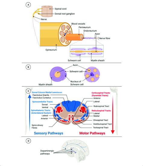
Table of Contents
Bundles of Axons Within the Central Nervous System are Called Tracts: A Deep Dive into Neurological Structure and Function
Bundles of axons within the central nervous system (CNS) are called tracts. Understanding tracts is crucial to comprehending the complex communication network that allows our brains and spinal cords to function. This article will delve deep into the anatomy, function, and clinical significance of these crucial neurological structures. We'll explore different types of tracts, their classification, and the consequences of damage to these vital pathways.
What are Tracts? A Definition and Overview
Tracts are collections of nerve fibers (axons) that share a common origin, destination, and function within the CNS. Unlike nerves in the peripheral nervous system (PNS), which are bundles of both axons and dendrites, tracts consist exclusively of axons. These axons are often myelinated, increasing the speed of signal transmission. The myelin sheath, a fatty substance produced by oligodendrocytes in the CNS, acts as insulation, ensuring efficient signal propagation along the axon. The arrangement and organization of these tracts are critical for the efficient processing and transmission of neural information throughout the brain and spinal cord.
Distinguishing Tracts from Nerves
It's important to differentiate between tracts (CNS) and nerves (PNS). This distinction lies primarily in their composition and location:
- Tracts: Found within the brain and spinal cord (CNS); composed solely of axons; myelinated by oligodendrocytes.
- Nerves: Found outside the brain and spinal cord (PNS); composed of both axons and dendrites; myelinated by Schwann cells.
This fundamental difference highlights the distinct functional roles and structural organization of the CNS and PNS.
Classification of Tracts Based on Function
Tracts are categorized based on the type of information they transmit. This functional classification is crucial for understanding the intricate pathways that govern sensory perception, motor control, and higher-order cognitive functions.
1. Sensory Tracts (Afferent Tracts)
Sensory tracts carry sensory information from the body to the brain. These pathways are essential for our perception of the external and internal environments. Examples include:
- Dorsal Column-Medial Lemniscus Pathway: Transmits fine touch, proprioception (sense of body position), and vibration.
- Spinothalamic Tract: Carries information about pain, temperature, and crude touch.
- Spinocerebellar Tracts: Transmit proprioceptive information to the cerebellum, crucial for coordination and balance.
These sensory tracts have specific pathways and relay stations within the CNS, allowing for the precise processing and interpretation of sensory stimuli.
2. Motor Tracts (Efferent Tracts)
Motor tracts carry motor commands from the brain to the muscles and glands. They are responsible for voluntary and involuntary movements. Key examples include:
- Corticospinal Tract (Pyramidal Tract): The major pathway for voluntary motor control, originating in the motor cortex and descending to the spinal cord. This tract is responsible for skilled, fine motor movements.
- Rubrospinal Tract: Involved in motor coordination and muscle tone, particularly in upper limb movements.
- Vestibulospinal Tract: Plays a role in maintaining balance and posture.
- Tectospinal Tract: Mediates head and eye movements in response to visual stimuli.
3. Association Tracts
Association tracts connect different areas within the same hemisphere of the brain. These tracts are essential for integrating information from various cortical regions and facilitating complex cognitive functions. Examples include:
- Arcuate Fasciculus: Connects Wernicke's area (language comprehension) to Broca's area (speech production), crucial for fluent speech.
- Cingulum: Connects the frontal lobe with the temporal and parietal lobes, involved in memory and emotional processing.
- Uncinate Fasciculus: Connects the frontal and temporal lobes, implicated in semantic memory and social cognition.
4. Commissural Tracts
Commissural tracts connect corresponding areas in the two hemispheres of the brain. The most prominent example is:
- Corpus Callosum: The largest white matter structure in the brain, connecting the left and right cerebral hemispheres. It facilitates interhemispheric communication and coordination of functions.
Anatomy of Tracts: Myelin, Oligodendrocytes, and White Matter
The structural integrity of tracts is largely determined by the presence of myelin and the supporting cells that produce it.
Myelin Sheath
The myelin sheath, a fatty insulating layer surrounding many axons, is crucial for efficient signal transmission. In the CNS, oligodendrocytes are the myelin-producing cells. Each oligodendrocyte can myelinate multiple axons, unlike Schwann cells in the PNS, which myelinate only a single axon.
Oligodendrocytes
These glial cells are responsible for the formation and maintenance of the myelin sheath around axons within the CNS. Their proper functioning is essential for the rapid and efficient transmission of nerve impulses. Damage to oligodendrocytes can lead to demyelination, resulting in neurological dysfunction.
White Matter
Tracts are primarily composed of white matter, a tissue that gets its characteristic color from the myelin sheaths surrounding the axons. The organization of white matter tracts is highly complex and varies across different regions of the brain and spinal cord. The arrangement of these tracts reflects the intricate network of connections that allows for efficient information processing and communication.
Clinical Significance of Tract Damage
Damage to tracts can have significant neurological consequences, depending on the location and extent of the injury. Causes of tract damage include:
- Stroke (Cerebrovascular Accident): Disruption of blood supply to the brain can lead to damage or death of neurons and their axons.
- Traumatic Brain Injury (TBI): Physical trauma to the brain can result in damage to white matter tracts, causing a variety of neurological deficits.
- Multiple Sclerosis (MS): An autoimmune disease characterized by demyelination of axons in the CNS.
- Tumors: Tumors within the CNS can compress or invade white matter tracts, disrupting their function.
The clinical manifestations of tract damage vary greatly depending on the specific tract affected. Damage to sensory tracts can lead to sensory loss (e.g., loss of touch, pain, temperature sensation). Damage to motor tracts can result in weakness, paralysis, or incoordination. Damage to association or commissural tracts can cause a range of cognitive deficits, depending on the specific area affected.
Diagnostic Methods for Assessing Tract Integrity
Several techniques are used to assess the integrity of white matter tracts:
- Magnetic Resonance Imaging (MRI): MRI provides high-resolution images of brain structures, allowing visualization of white matter tracts. Diffusion tensor imaging (DTI), a type of MRI, is particularly useful for assessing the microstructure of white matter.
- Diffusion Tensor Imaging (DTI): A specialized MRI technique that measures the diffusion of water molecules along white matter tracts. It allows for the reconstruction of 3D images of white matter pathways and the assessment of their integrity.
- Tractography: A technique that uses DTI data to reconstruct the three-dimensional pathways of white matter tracts. This allows for visualization of the connections between different brain regions.
Research and Future Directions
Ongoing research continues to explore the complexities of white matter tracts and their role in various neurological functions. Advances in neuroimaging techniques are providing increasingly detailed insights into the structure and function of these pathways. Future research may focus on:
- Developing more refined techniques for imaging and analyzing white matter tracts.
- Investigating the role of white matter integrity in various neurological and psychiatric disorders.
- Exploring the potential for therapeutic interventions to repair damaged white matter tracts.
Conclusion
Bundles of axons within the central nervous system are called tracts. These crucial structures form the intricate communication network that allows our brains and spinal cords to function. Understanding their anatomy, function, and clinical significance is essential for comprehending the complexities of the nervous system and for diagnosing and treating neurological disorders. Ongoing research continues to unravel the mysteries of these vital pathways, offering hope for improved diagnostic and therapeutic strategies in the future. The diverse types of tracts, their intricate organization, and the devastating consequences of damage highlight their critical role in maintaining neurological health and overall well-being. Further exploration into the intricacies of these pathways will undoubtedly lead to advancements in our understanding of the brain and the treatment of neurological diseases.
Latest Posts
Latest Posts
-
Is Trp Operon Inducible Or Repressible
Mar 29, 2025
-
As A Result Of A Decrease In Price
Mar 29, 2025
-
The Nuclear Membrane Reappears In Mitosis During
Mar 29, 2025
-
How Much Seconds Are In A Week
Mar 29, 2025
-
Is Boron A Gas Solid Or Liquid
Mar 29, 2025
Related Post
Thank you for visiting our website which covers about Bundles Of Axons Within The Central Nervous System Are Called . We hope the information provided has been useful to you. Feel free to contact us if you have any questions or need further assistance. See you next time and don't miss to bookmark.
