A Joint Between Bones Of The Skull Is A
News Leon
Apr 06, 2025 · 7 min read
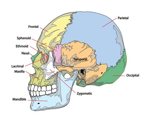
Table of Contents
A Joint Between Bones of the Skull Is a Suture: A Deep Dive into Cranial Anatomy
The human skull, a marvel of engineering, is composed of numerous bones intricately joined together to form a protective vault for the brain. Unlike the freely moving joints found in the limbs, the connections between skull bones are immovable fibrous joints known as sutures. Understanding the anatomy, function, and clinical significance of these sutures is crucial for anyone studying anatomy, medicine, or related fields. This comprehensive article will explore the intricacies of sutures, their classification, developmental aspects, clinical relevance, and more.
What is a Suture?
A suture is a type of fibrous joint specifically found in the skull. Fibrous joints are characterized by the presence of fibrous connective tissue connecting the bones. Unlike other fibrous joints like gomphoses (teeth in sockets) or syndesmoses (slightly moveable joints like the distal tibiofibular joint), sutures are virtually immobile, designed to provide strong, interlocking connections that protect the brain. This immobility is essential for safeguarding the delicate brain tissue from external forces. The bones are held together by a thin layer of dense, fibrous connective tissue rich in collagen fibers. This dense tissue contributes to the remarkable strength and stability of the skull.
Types of Sutures: A Detailed Look
Sutures are not all created equal. They are classified based on the shape of the interlocking bony edges:
-
Serrate Sutures: These are the most common type, characterized by interlocking, saw-tooth edges. This interlocking design significantly enhances the strength and stability of the joint. Examples include the sagittal suture (between the two parietal bones) and the coronal suture (between the frontal and parietal bones). The intricate interdigitation of the serrate suture significantly increases the surface area of contact between the bones, making them highly resistant to shearing forces.
-
Squamous Sutures: These sutures are characterized by overlapping, beveled edges. The bones appear to overlap each other like shingles on a roof. A prime example is the squamous suture between the temporal and parietal bones. The beveled edges create a strong, yet relatively smooth articulation.
-
Plane Sutures: These sutures exhibit relatively straight, non-overlapping edges. They are less common than serrate or squamous sutures. An example is the intermaxillary suture found between the palatine processes of the maxillae. While seemingly less intricate, these sutures still provide a sturdy connection.
-
Dentoalveolar Syndesmoses (Gomphoses): While technically a separate category of fibrous joint, dentoalveolar syndesmoses, commonly known as gomphoses, merit mention here due to their close relationship to the skull. These are the peg-in-socket joints found between the teeth and the alveolar processes of the maxilla and mandible. The fibrous connective tissue in this case is the periodontal ligament.
Sutural Development and Ossification
The development of sutures is an intricate process linked to the overall development of the skull. During fetal development, the skull is formed from several separate bones, which gradually grow and fuse together through the process of intramembranous ossification. In this process, mesenchymal cells differentiate directly into osteoblasts, which then secrete bone matrix. This process forms the flat bones of the skull. The sutures themselves are formed from the remaining undifferentiated mesenchymal tissue between the ossifying bones.
As the child grows, the sutures remain open allowing for the expansion of the skull to accommodate the growing brain. This flexibility is crucial for facilitating the brain's rapid development during infancy and childhood. The degree of ossification varies greatly depending on the suture and individual variability. It's important to note that the degree of fusion may not be uniform across all sutures even in adults.
The gradual ossification of the sutures is a continuous process that typically continues well into adulthood. However, the timing and extent of fusion can vary between individuals, and some sutures may remain patent (open) for a longer period or even throughout life. This phenomenon is thought to play a role in accommodating slight changes in skull shape throughout life and potentially contribute to skull flexibility under stress.
Clinical Significance of Sutures
While sutures primarily serve a protective function, their clinical significance extends beyond simple bone connection. Several conditions are directly or indirectly linked to sutural abnormalities:
-
Craniosynostosis: This is a condition characterized by premature fusion of one or more sutures. This can lead to abnormal skull shape and potentially compromise brain development, necessitating surgical intervention. The severity of craniosynostosis depends on the affected suture(s) and the extent of premature fusion. For example, premature closure of the sagittal suture can lead to a long, narrow skull (scaphocephaly), while premature closure of the coronal suture can result in a short, wide skull (brachycephaly).
-
Skull Fractures: Sutures can be sites of skull fractures. The complex interlocking nature of many sutures can lead to complex fracture patterns. The proximity to the brain makes skull fractures particularly concerning. Careful assessment and management are crucial to minimize the risk of brain injury.
-
Infections: Sutures can serve as potential pathways for the spread of infection from the scalp to the brain. This highlights the importance of proper management of scalp injuries and infections. The close proximity of the meninges to the sutures necessitates swift and effective treatment of infections in this area.
-
Age-Related Changes: As people age, the sutures undergo progressive ossification and fusion. This process is largely asymptomatic but might be visible on radiographic imaging. The timing and extent of ossification vary greatly depending on genetic and environmental factors.
-
Surgical Considerations: Neurosurgeons must carefully consider the location and anatomy of sutures during cranial surgeries. Precise knowledge of suture location is vital to minimize damage during surgical procedures.
Imaging Techniques for Studying Sutures
Several imaging techniques allow visualization of sutures and assess their integrity:
-
Plain X-rays: While not providing the detailed anatomical information of other imaging modalities, plain radiographs are often the initial imaging study used for the assessment of skull fractures and sutural fusion. They can provide information on overall skull shape and detect obvious abnormalities.
-
Computed Tomography (CT): CT scans offer detailed cross-sectional images of the skull, providing superior visualization of the sutures and bony structures. This is invaluable for assessing skull fractures, craniosynostosis, and other sutural abnormalities. The high resolution of CT allows for precise assessment of fracture lines and their relationship to sutures.
-
Magnetic Resonance Imaging (MRI): Although primarily used for visualizing soft tissues, MRI can also provide complementary information regarding the relationship of the sutures to surrounding brain structures, especially in cases of craniosynostosis. MRI can help assess potential complications like brain herniation associated with premature sutural fusion.
The Significance of Understanding Sutures
Understanding the anatomy, development, and clinical significance of sutures is crucial for numerous healthcare professionals. Neurosurgeons need precise knowledge of suture locations for precise surgical interventions. Radiologists must interpret imaging studies accurately to identify sutural abnormalities. Pediatricians need to recognize the signs and symptoms of craniosynostosis. Moreover, anthropologists and forensic scientists can utilize suture patterns to determine age and sex in skeletal remains. The study of sutures provides a window into human development, evolution, and disease processes. The intricate interlocking nature of these joints and their crucial role in protecting the delicate brain highlights the importance of understanding their unique characteristics.
Conclusion
The joints between bones of the skull, known as sutures, are remarkably specialized fibrous joints designed for both strength and flexibility. Their intricate morphology, development, and clinical relevance continue to fascinate and challenge scientists and clinicians alike. From their role in protecting the brain to their involvement in various pathologies, sutures represent a vital area of study across multiple disciplines. Continued research in this area will undoubtedly deepen our understanding of human biology and contribute to improved diagnosis and treatment of conditions affecting the skull. Appreciating the complexities of these seemingly simple structures allows for a more thorough understanding of human anatomy and the intricacies of cranial development and physiology.
Latest Posts
Latest Posts
-
Osmosis Is A Type Of Active Transport
Apr 08, 2025
-
What Is The Major Product Of This Reaction
Apr 08, 2025
-
Which Of The Following Is An Example Of Artificial Selection
Apr 08, 2025
-
How To Write A Matrix In Python
Apr 08, 2025
-
What Is The Largest Phylum Of Invertebrates
Apr 08, 2025
Related Post
Thank you for visiting our website which covers about A Joint Between Bones Of The Skull Is A . We hope the information provided has been useful to you. Feel free to contact us if you have any questions or need further assistance. See you next time and don't miss to bookmark.