Which Of The Following Bones Belong To The Axial Skeleton
News Leon
Mar 23, 2025 · 6 min read
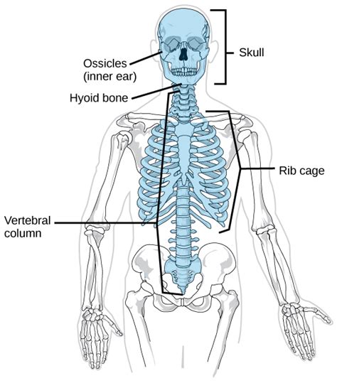
Table of Contents
Which Bones Belong to the Axial Skeleton? A Comprehensive Guide
The human skeleton is a marvel of engineering, providing structure, support, and protection for our bodies. It's broadly divided into two main sections: the axial skeleton and the appendicular skeleton. Understanding the difference is crucial for anyone studying anatomy, physiology, or simply curious about the human body. This article will delve deep into the axial skeleton, identifying its constituent bones and their crucial roles.
What is the Axial Skeleton?
The axial skeleton forms the central axis of the body. Think of it as the core structure upon which the limbs are attached. It's comprised of 80 bones, and its primary function is to protect vital organs and provide support for the head, neck, and trunk. This central framework allows for movement and stability, enabling essential functions like breathing and posture maintenance.
The Bones of the Axial Skeleton: A Detailed Breakdown
Let's break down the axial skeleton bone by bone, categorized for clarity:
1. The Skull (Cranium and Facial Bones):
The skull, a complex structure, protects the brain, the body's control center. It's divided into two main parts:
-
Cranial Bones: These eight bones form the protective cranial cavity. They include:
- Frontal Bone: Forms the forehead and upper eye sockets.
- Parietal Bones (2): Form the sides and roof of the cranium.
- Temporal Bones (2): Located on the sides of the skull, housing the inner ear and the temporomandibular joint (TMJ).
- Occipital Bone: Forms the back of the skull, containing the foramen magnum, the large opening where the spinal cord connects to the brain.
- Sphenoid Bone: A complex, bat-shaped bone located at the base of the skull, crucial for cranial support and muscle attachment.
- Ethmoid Bone: A delicate bone forming part of the nasal cavity and eye sockets.
-
Facial Bones: These fourteen bones contribute to the structure of the face. Key bones include:
- Nasal Bones (2): Form the bridge of the nose.
- Maxillae (2): Form the upper jaw, holding the upper teeth.
- Zygomatic Bones (2): Form the cheekbones.
- Mandible: The lower jawbone, the only movable bone in the skull.
- Lacrimal Bones (2): Small bones forming part of the medial wall of each orbit (eye socket).
- Palatine Bones (2): Form the posterior part of the hard palate (roof of the mouth).
- Inferior Nasal Conchae (2): Scroll-like bones within the nasal cavity, increasing its surface area.
- Vomer: A single, flat bone forming part of the nasal septum (the partition separating the nostrils).
2. The Vertebral Column (Spine):
The vertebral column, also known as the spine or backbone, is a flexible column of 33 vertebrae. It provides structural support, protects the spinal cord, and enables movement. It's divided into five regions:
-
Cervical Vertebrae (7): The seven vertebrae in the neck, characterized by their smaller size and transverse foramina (holes in the transverse processes). The atlas (C1) and axis (C2) are unique, allowing for head rotation and nodding.
-
Thoracic Vertebrae (12): These vertebrae articulate with the ribs, contributing to the thoracic cage's structure. They are larger than the cervical vertebrae and have long, downward-sloping spinous processes.
-
Lumbar Vertebrae (5): The five lumbar vertebrae in the lower back are the largest and strongest vertebrae, supporting most of the body's weight.
-
Sacral Vertebrae (5): These five vertebrae are fused together to form the sacrum, a triangular bone that forms the posterior wall of the pelvis.
-
Coccygeal Vertebrae (4): These four vertebrae are fused to form the coccyx (tailbone), a vestigial structure representing the remnant of a tail.
3. The Thoracic Cage (Rib Cage):
The thoracic cage, also known as the rib cage, protects vital organs like the heart and lungs. It's composed of:
-
Sternum: The breastbone, a flat bone located in the anterior chest wall. It's comprised of three parts: the manubrium (upper part), the body (middle part), and the xiphoid process (lower part).
-
Ribs (24): Twelve pairs of ribs arching around the chest cavity. The first seven pairs are true ribs, directly articulating with the sternum via costal cartilage. The next three pairs are false ribs, indirectly attached to the sternum through the costal cartilage of the seventh rib. The last two pairs are floating ribs, lacking any anterior attachment to the sternum.
The Importance of Understanding the Axial Skeleton
Knowing the bones of the axial skeleton is essential for various reasons:
-
Medical Diagnosis: Accurate identification of bones is crucial for diagnosing injuries and diseases affecting the skull, spine, or rib cage. Conditions like fractures, scoliosis, and spinal stenosis can be diagnosed and managed effectively only with a thorough understanding of the anatomy.
-
Surgical Procedures: Surgeons require detailed knowledge of the axial skeleton for planning and executing complex procedures like spinal surgeries, craniofacial surgeries, and chest surgeries. Precise anatomical understanding minimizes risks and improves surgical outcomes.
-
Physical Therapy and Rehabilitation: Physical therapists use their knowledge of the axial skeleton to develop rehabilitation programs for patients recovering from injuries or surgeries. They focus on restoring mobility, strength, and function.
-
Forensic Science: Forensic scientists use their knowledge of skeletal anatomy, including the axial skeleton, to identify victims and determine the cause of death in criminal investigations.
-
Artistic Anatomy: Artists, especially those involved in anatomical illustration, need a strong understanding of the axial skeleton to realistically depict the human form. This knowledge helps them to portray the skeletal structure's accurate proportions, relationships, and movements.
Beyond the Bones: Connections and Function
The axial skeleton isn't just a collection of individual bones. Its intricate structure relies on:
-
Joints: These are where bones meet, enabling movement and providing stability. The skull's joints are largely immovable (fibrous), while the vertebrae are joined by flexible intervertebral discs. The rib cage demonstrates a combination of cartilaginous and synovial joints.
-
Ligaments: Strong, fibrous tissues connecting bones together at joints, providing stability and preventing excessive movement. Ligaments are vital in maintaining the integrity of the spine and rib cage.
-
Muscles: Muscles are attached to bones, enabling movement of the head, neck, and trunk. The complex interplay of muscles and bones facilitates diverse actions like breathing, posture maintenance, and head and neck movements.
Common Conditions Affecting the Axial Skeleton
Several conditions can affect the bones of the axial skeleton, including:
-
Scoliosis: A sideways curvature of the spine.
-
Kyphosis: An excessive outward curvature of the thoracic spine (hunchback).
-
Lordosis: An excessive inward curvature of the lumbar spine (swayback).
-
Osteoporosis: A bone disease characterized by low bone mass and structural deterioration, leading to increased fracture risk.
-
Spinal Stenosis: A narrowing of the spinal canal, causing pressure on the spinal cord or nerves.
Conclusion
The axial skeleton is a fundamental component of the human body, providing structural support, protection for vital organs, and facilitating essential movements. Its complex arrangement of bones, joints, ligaments, and muscles demonstrates remarkable engineering and biological integration. Understanding its anatomy, functions, and common associated conditions is crucial for various healthcare professions and those curious about the intricacies of the human body. This detailed breakdown offers a comprehensive overview, paving the way for deeper exploration and a more profound appreciation of this remarkable structure. Remember to consult medical professionals for any concerns regarding your skeletal health.
Latest Posts
Latest Posts
-
The Product Of Two Irrrational Numbers Is Irrational
Mar 24, 2025
-
What Is The Square Root Of 4761
Mar 24, 2025
-
A Square Formed By Four Isosceles Triangles
Mar 24, 2025
-
Which Tunic Of An Artery Contains Endothelium
Mar 24, 2025
-
How Many Minutes Are In 60 Hours
Mar 24, 2025
Related Post
Thank you for visiting our website which covers about Which Of The Following Bones Belong To The Axial Skeleton . We hope the information provided has been useful to you. Feel free to contact us if you have any questions or need further assistance. See you next time and don't miss to bookmark.
