Which Chamber Has The Thickest Muscular Wall
News Leon
Mar 24, 2025 · 6 min read
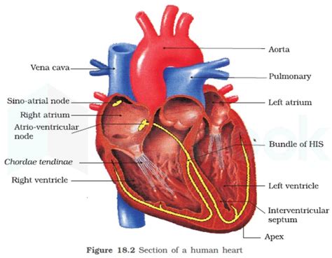
Table of Contents
Which Chamber Has the Thickest Muscular Wall? Understanding the Heart's Powerful Anatomy
The human heart, a tireless engine driving our circulatory system, is a marvel of biological engineering. Composed of four chambers – two atria and two ventricles – each plays a crucial role in pumping blood throughout the body. But one key difference sets these chambers apart: the thickness of their muscular walls. Understanding why this variation exists is key to grasping the heart's functional mechanics and appreciating its remarkable adaptability. This article delves deep into the anatomical structure of the heart, explaining why the left ventricle boasts the thickest muscular wall, and the vital implications of this anatomical feature.
The Heart's Chambers: A Functional Overview
Before diving into the specifics of muscular wall thickness, let's briefly review the roles of each heart chamber:
The Atria: Receiving Chambers
The two atria, the right atrium and the left atrium, are the heart's receiving chambers. They receive blood returning to the heart from the body (right atrium) and the lungs (left atrium). Their muscular walls are relatively thin because their function is primarily to passively collect blood and then pump it into the ventricles. This gentle pumping action requires less muscular force compared to the ventricles.
The Ventricles: Powerful Pumping Chambers
The ventricles, the right ventricle and the left ventricle, are the heart's powerful pumping chambers. They receive blood from the atria and then forcefully eject it into the pulmonary artery (right ventricle) and the aorta (left ventricle), propelling blood to the lungs and the rest of the body respectively. The significant difference in the workload of these two ventricles directly correlates with the thickness of their muscular walls.
Why the Left Ventricle Has the Thickest Wall: The Pressure Factor
The left ventricle possesses the thickest muscular wall because it must generate significantly higher pressure than any other chamber. This higher pressure is absolutely necessary to overcome the systemic vascular resistance, the resistance the blood encounters as it flows through the vast network of blood vessels in the body. Think of it like this: the left ventricle needs to pump blood all the way to the toes, overcoming the friction and resistance along the way, a significantly longer and more challenging journey than the blood from the right ventricle travels to the lungs.
Several factors contribute to this pressure difference:
-
Systemic Circulation vs. Pulmonary Circulation: The systemic circulation (left ventricle to body) involves a much longer and more extensive network of blood vessels compared to the pulmonary circulation (right ventricle to lungs). This increased length and smaller diameter vessels contribute to higher resistance to blood flow.
-
Blood Pressure: The systemic circulation operates under significantly higher blood pressure than the pulmonary circulation. The left ventricle must generate enough force to maintain this higher pressure, requiring a thicker muscular wall.
-
Blood Volume: The volume of blood pumped by the left ventricle is comparable to the right ventricle. However, the much higher pressure required by the systemic circulation demands that the left ventricle exert considerably more force with each contraction.
Microscopic Anatomy: Understanding the Muscular Structure
The increased thickness of the left ventricle's wall is not just about sheer mass; it's also about the organization and type of muscle fibers. The walls of the ventricles are composed primarily of cardiac muscle, specialized muscle tissue that enables rhythmic contractions. The left ventricle’s wall contains a higher density of cardiomyocytes (heart muscle cells) arranged in a complex, interwoven pattern that allows for efficient force generation and coordinated contraction. This intricate arrangement optimizes the force of ejection, further emphasizing the ventricle’s crucial role in systemic circulation.
Analyzing the microscopic structure reveals a more complex picture:
-
Myocardial Fibers: The left ventricle's wall is composed of layers of myocardial fibers, spiraling around the chamber in a complex pattern. This arrangement optimizes force transmission during contraction, ensuring efficient ejection of blood into the aorta.
-
Cardiac Muscle Cells: The individual cardiac muscle cells (cardiomyocytes) in the left ventricle are larger and more densely packed than those in the other chambers, further enhancing the force of contraction.
-
Connective Tissue: The intricate arrangement of muscle fibers is supported by a robust network of connective tissue, providing structural integrity and preventing overstretching during contraction.
Comparing the Ventricular Walls: A Detailed Analysis
Let's compare the left and right ventricle's wall thickness more directly:
| Chamber | Wall Thickness (Approximate) | Functional Role | Pressure Generated |
|---|---|---|---|
| Left Ventricle | 1-1.5 cm | Pumps oxygenated blood to the body | High pressure, overcoming systemic vascular resistance |
| Right Ventricle | 0.5-0.7 cm | Pumps deoxygenated blood to the lungs | Low pressure |
The significant difference in wall thickness clearly reflects the distinct demands of systemic versus pulmonary circulation. The left ventricle's thicker wall allows it to generate the high pressure necessary for efficient blood flow throughout the body, while the right ventricle's thinner wall is perfectly suited for the lower-pressure requirements of pulmonary circulation.
Clinical Implications: Understanding Heart Disease
The varying thickness of the heart chambers has important clinical implications. Conditions like hypertrophy, an increase in the size of the heart muscle, often manifest most prominently in the left ventricle. This is because the left ventricle works harder than the right ventricle, making it more susceptible to strain and enlargement in response to various cardiovascular diseases.
Understanding the left ventricle's anatomy is crucial for diagnosing and managing various heart conditions, including:
-
Hypertrophic Cardiomyopathy (HCM): HCM is characterized by an abnormal thickening of the left ventricular wall, which can impair the heart's ability to pump blood effectively.
-
Dilated Cardiomyopathy (DCM): DCM involves a thinning and weakening of the left ventricular wall, leading to impaired pumping ability.
-
Coronary Artery Disease (CAD): Reduced blood flow to the left ventricle due to CAD can significantly impair its function and lead to heart failure.
Conclusion: Appreciating the Heart's Adaptive Power
The fact that the left ventricle has the thickest muscular wall is a testament to the remarkable adaptability of the human heart. This anatomical feature is not a random occurrence but a direct consequence of the different physiological demands placed on the left and right ventricles. The left ventricle's robust muscular structure enables it to overcome the high resistance of systemic circulation, ensuring efficient delivery of oxygenated blood to all parts of the body. A deeper understanding of this vital difference allows for a greater appreciation of the heart's intricate design and its critical role in maintaining our overall health. Further research into the specific cellular mechanisms and genetic influences shaping ventricular wall thickness continues to be a crucial area of cardiovascular research. The more we understand about this fundamental aspect of cardiac anatomy, the better equipped we will be to diagnose, treat, and prevent heart disease.
Latest Posts
Related Post
Thank you for visiting our website which covers about Which Chamber Has The Thickest Muscular Wall . We hope the information provided has been useful to you. Feel free to contact us if you have any questions or need further assistance. See you next time and don't miss to bookmark.
