When The Diaphragm And External Intercostal Muscles Contract
News Leon
Mar 15, 2025 · 5 min read
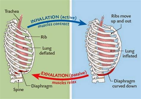
Table of Contents
When the Diaphragm and External Intercostal Muscles Contract: A Deep Dive into Inspiration
Breathing, a seemingly effortless act, is a complex interplay of muscular contractions and pressure changes. Understanding the mechanics of inspiration, the process of inhaling, requires a detailed look at the primary muscles involved: the diaphragm and the external intercostal muscles. When these muscles contract, they initiate a cascade of events leading to the inflow of air into the lungs. This article will delve deep into this process, exploring the anatomy, physiology, and implications of their coordinated action.
The Anatomy of Inspiration: Players on the Stage
Before understanding the mechanics of contraction, let's examine the key players:
The Diaphragm: The Primary Muscle of Inspiration
The diaphragm, a dome-shaped muscle separating the thoracic (chest) cavity from the abdominal cavity, is the most important muscle involved in quiet breathing. Its unique structure is crucial to its function. Imagine it as a parachute: a large, thin sheet of muscle with a central tendon.
- Origin: The diaphragm originates from the xiphoid process of the sternum (breastbone), the lower six ribs, and the lumbar vertebrae (lower back bones).
- Insertion: All these muscle fibers converge and insert into the central tendon.
- Innervation: The phrenic nerve, originating from cervical spinal nerves C3-C5, innervates the diaphragm. This is crucial as damage to these nerves can severely impair breathing.
The External Intercostal Muscles: Supporting the Star
The external intercostal muscles, eleven pairs of muscles located between the ribs, play a crucial supporting role in inspiration. They run obliquely, from the lower border of a rib to the upper border of the rib below.
- Origin: The lower border of a rib.
- Insertion: The upper border of the rib below.
- Innervation: Intercostal nerves, branches of the thoracic spinal nerves, supply the external intercostals.
The Mechanics of Contraction: A Symphony of Movement
When we breathe in, a coordinated contraction of the diaphragm and external intercostal muscles occurs:
Diaphragm Contraction: The Expanding Dome
When the phrenic nerve sends signals, the diaphragm contracts. This causes the dome-shaped diaphragm to flatten, increasing the vertical dimension of the thoracic cavity. Think of it like pushing down on the dome of a parachute; it flattens out, increasing the space beneath it. This downward movement is substantial, contributing significantly to lung volume increase.
External Intercostal Muscle Contraction: The Rib Cage Expansion
Simultaneously, the external intercostal muscles contract. This contraction elevates the ribs and expands the chest laterally (sideways) and anteroposteriorly (front to back). The ribs rotate upward and outward, increasing the transverse and anteroposterior dimensions of the thoracic cavity. This action further increases the overall volume of the chest.
Pressure Changes: The Driving Force of Inspiration
These muscular contractions create a chain reaction of pressure changes that drive air into the lungs:
Decrease in Intrapleural Pressure: The Crucial Step
The expansion of the thoracic cavity leads to a decrease in intrapleural pressure—the pressure within the pleural space, the space between the lungs and the chest wall. This negative pressure creates a pressure gradient, pulling the lungs outwards to expand.
Decrease in Intrapulmonary Pressure: Drawing Air Inward
The expansion of the lungs leads to a decrease in intrapulmonary pressure—the pressure within the alveoli (tiny air sacs in the lungs). This lower pressure within the lungs is now less than the atmospheric pressure outside, creating a pressure gradient that draws air into the lungs until the pressures equalize.
Beyond Quiet Breathing: Forced Inspiration and Accessory Muscles
While the diaphragm and external intercostals are sufficient for quiet breathing, forced inspiration, such as during exercise or strenuous activity, requires the recruitment of accessory muscles. These include:
- Sternocleidomastoid: Elevates the sternum and increases the anteroposterior diameter of the chest.
- Scalenes: Elevate the first two ribs.
- Pectoralis minor: Elevates the ribs.
- Serratus anterior: Elevates the ribs.
These muscles augment the action of the diaphragm and external intercostals, further increasing the volume of the thoracic cavity and enabling a deeper inhalation.
Clinical Significance: Understanding Breathing Disorders
Understanding the mechanics of diaphragm and external intercostal muscle contraction is essential for diagnosing and treating various respiratory disorders. Conditions affecting these muscles can significantly impair breathing, leading to:
- Respiratory distress: Difficulty breathing, often characterized by shortness of breath and rapid breathing.
- Hypoxemia: Low blood oxygen levels, potentially leading to organ damage and even death.
- Hypercapnia: Elevated carbon dioxide levels in the blood, which can cause acid-base imbalances and neurological dysfunction.
Conditions that can affect these muscles include:
- Diaphragmatic paralysis: Weakness or paralysis of the diaphragm, often due to nerve damage.
- Muscular dystrophies: Genetic disorders causing progressive muscle weakness and wasting.
- Respiratory infections: Infections like pneumonia can inflame the lungs and chest wall, hindering the effective contraction of respiratory muscles.
- Trauma: Chest injuries can damage the ribs, intercostal muscles, or diaphragm, compromising breathing mechanics.
Optimizing Respiratory Function: Exercise and Lifestyle
Maintaining the health and strength of the diaphragm and intercostal muscles is crucial for optimal respiratory function. Several strategies can help:
- Diaphragmatic breathing exercises: These exercises help strengthen the diaphragm and improve breathing efficiency.
- Regular exercise: Cardiovascular exercise improves overall fitness and strengthens respiratory muscles.
- Good posture: Maintaining good posture ensures optimal positioning of the chest and ribs, allowing for efficient respiratory muscle function.
- Avoiding smoking: Smoking damages the lungs and respiratory muscles, impairing breathing.
The Interplay of Muscles: A Complex but Efficient System
The coordination between the diaphragm and external intercostal muscles is a remarkable example of the body's intricate design. Their precisely timed contractions create the pressure changes necessary for efficient air exchange. Understanding this mechanism is crucial for appreciating the complexity of respiration and for diagnosing and managing respiratory disorders. Furthermore, practicing techniques to strengthen these key muscles can contribute significantly to overall respiratory health and well-being.
Conclusion: Breathing – More Than Just Inhaling and Exhaling
Breathing is far more intricate than the simple act of inhaling and exhaling. The coordinated contractions of the diaphragm and external intercostal muscles, coupled with the resulting pressure changes, form the foundation of this vital process. This detailed look reveals the crucial roles of these muscles and highlights the importance of maintaining their health and functionality for optimal respiratory health and overall well-being. Understanding this process empowers individuals to take proactive steps towards improving their respiratory function and overall quality of life. The more we know about the mechanics of breathing, the better equipped we are to protect and enhance this fundamental life function.
Latest Posts
Related Post
Thank you for visiting our website which covers about When The Diaphragm And External Intercostal Muscles Contract . We hope the information provided has been useful to you. Feel free to contact us if you have any questions or need further assistance. See you next time and don't miss to bookmark.
