What Serous Membrane Covers The Lungs
News Leon
Apr 06, 2025 · 6 min read
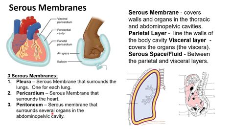
Table of Contents
What Serous Membrane Covers the Lungs? Understanding the Pleura
The lungs, essential organs for respiration, are not simply floating freely within the thoracic cavity. Instead, they are enveloped and protected by a delicate yet crucial serous membrane known as the pleura. Understanding the pleura's structure, function, and clinical significance is paramount to grasping the intricacies of pulmonary physiology and pathology. This comprehensive article will delve deep into the anatomy and physiology of the pleura, exploring its layers, functions, and the implications of pleural abnormalities.
The Pleura: A Protective Embrace
The pleura is a thin, double-layered serous membrane that completely encloses each lung. Think of it as a specialized, airtight sac that creates a unique environment for the lungs to expand and contract efficiently. This crucial membrane is comprised of two distinct layers:
1. Visceral Pleura: The Lung's Intimate Lining
The visceral pleura is the innermost layer, intimately adhered to the surface of each lung. It follows every contour, fissure, and lobe of the lung, essentially acting as a second skin. This close apposition ensures that the pleura moves with the lung during respiration, facilitating its expansion and contraction. The visceral pleura is richly supplied with nerve endings, contributing to the sensory innervation of the lung itself, although the lung tissue is relatively insensitive to pain.
2. Parietal Pleura: Anchoring the Lung to the Thoracic Cavity
The parietal pleura is the outer layer, lining the thoracic cavity. Unlike the visceral pleura, it is not directly attached to the lung. Instead, it lines the inner surface of the chest wall, the diaphragm (the floor of the thoracic cavity), the mediastinum (the central compartment of the chest containing the heart, great vessels, and esophagus), and the superior surface of the diaphragm. This anchoring role is vital for maintaining the lung's position and preventing its collapse.
The Pleural Cavity: A Potential Space with Significant Implications
Between the visceral and parietal pleura lies the pleural cavity. Crucially, this is not an empty space; rather, it's a potential space containing a small amount of pleural fluid. This fluid acts as a lubricant, minimizing friction between the two pleural layers as the lungs expand and contract during breathing. The thin layer of fluid creates surface tension, holding the visceral and parietal pleura together, ensuring that the lung moves in unison with the chest wall. The negative pressure within the pleural cavity is essential for lung inflation and overall respiratory function. This negative pressure, or intrapleural pressure, is lower than both atmospheric pressure and intrapulmonary pressure (the pressure inside the lungs).
The Pleura's Vital Functions
The pleura's role extends far beyond simple physical protection. Its functions are multifaceted and essential for normal respiratory mechanics:
-
Lubrication and Reduced Friction: The pleural fluid acts as an effective lubricant, reducing friction between the moving surfaces of the visceral and parietal pleura during respiration. This minimizes energy expenditure and prevents damage to the delicate lung tissue.
-
Lung Expansion and Contraction: The negative pressure within the pleural cavity facilitates lung inflation. As the diaphragm contracts and the chest wall expands, the intrapleural pressure becomes even more negative, drawing air into the lungs. During exhalation, the process reverses.
-
Compartmentalization: The pleura separates the lungs from other structures in the thoracic cavity, preventing the spread of infection or inflammation. This compartmentalization is a critical protective mechanism.
-
Pulmonary Compliance: The pleura contributes significantly to pulmonary compliance, the ease with which the lungs can expand and contract. A healthy pleura ensures optimal lung function.
Clinical Significance of Pleural Abnormalities
Several pathological conditions can affect the pleura, leading to significant respiratory compromise. Understanding these conditions is crucial for accurate diagnosis and appropriate management:
1. Pleurisy (Pleuritis): Inflammation of the Pleura
Pleurisy, or pleuritis, is an inflammation of the pleura. It's often characterized by sharp, stabbing chest pain, particularly during deep breaths or coughs. The pain arises from the inflammation irritating the nerve endings in the parietal pleura. Various factors can cause pleurisy, including infections (viral, bacterial, fungal), autoimmune diseases, cancer, and pulmonary embolism. The inflammation can lead to increased pleural fluid production (pleural effusion), further impairing respiratory function.
2. Pleural Effusion: Excess Fluid in the Pleural Cavity
Pleural effusion refers to the accumulation of excess fluid in the pleural cavity. This fluid can be transudative (due to systemic factors like heart failure) or exudative (due to inflammation or infection). The presence of pleural effusion can compress the lung, reducing its ability to expand and leading to shortness of breath and decreased oxygen levels. Treatment often involves removing the excess fluid through a procedure called thoracentesis.
3. Pneumothorax: Collapsed Lung
A pneumothorax is a condition where air enters the pleural cavity, causing the lung to collapse. This can occur spontaneously (spontaneous pneumothorax), due to trauma (traumatic pneumothorax), or as a complication of underlying lung disease (tension pneumothorax). The entry of air disrupts the negative pressure in the pleural cavity, leading to lung collapse and respiratory distress. Treatment often involves inserting a chest tube to remove the air and re-inflate the lung.
4. Mesothelioma: A Rare but Aggressive Cancer
Mesothelioma is a rare but aggressive cancer affecting the mesothelium, the lining of the body's internal organs, including the pleura. It's most commonly associated with exposure to asbestos. Mesothelioma is notoriously difficult to treat, and the prognosis is often poor.
5. Pleural Thickening: Scarring of the Pleura
Pleural thickening involves the scarring and stiffening of the pleura. This can occur as a result of repeated inflammation, infection, or exposure to harmful substances like asbestos. Pleural thickening can restrict lung expansion, leading to shortness of breath and reduced lung capacity.
Diagnostic Procedures for Pleural Conditions
Several diagnostic procedures can help identify and assess pleural abnormalities:
-
Chest X-ray: A chest X-ray is often the first imaging test used to evaluate pleural conditions. It can reveal the presence of pleural effusion, pneumothorax, or pleural thickening.
-
Computed Tomography (CT) Scan: A CT scan provides more detailed images of the chest, allowing for better visualization of pleural abnormalities.
-
Ultrasound: Ultrasound can be used to guide thoracentesis, a procedure to remove fluid from the pleural cavity. It can also help assess the characteristics of the pleural fluid.
-
Thoracentesis: This procedure involves inserting a needle into the pleural cavity to remove fluid for analysis. The fluid analysis can help determine the cause of pleural effusion.
-
Bronchoscopy: A bronchoscopy involves inserting a thin, flexible tube into the airways to visualize the lungs and collect samples. This procedure can be helpful in diagnosing underlying lung diseases that may be contributing to pleural abnormalities.
Conclusion: The Pleura's Unsung Importance
The pleura, a seemingly insignificant membrane, plays a vital and multifaceted role in respiratory health. Its intricate structure and functions are essential for efficient lung expansion and contraction, minimizing friction, and providing a protective barrier. Understanding the pleura's anatomy and the various pathologies that can affect it is crucial for clinicians to diagnose and manage a wide range of respiratory conditions, from simple pleurisy to the more aggressive mesothelioma. Further research into the complexities of pleural physiology and pathology will undoubtedly lead to improved diagnostic and therapeutic strategies for these often debilitating conditions. The seemingly simple structure of the pleura holds significant implications for the maintenance of respiratory health and overall well-being. Its subtle yet impactful role deserves further exploration and appreciation within the broader context of pulmonary function and disease.
Latest Posts
Latest Posts
-
What Structures Are Present Only In Animal Cells
Apr 07, 2025
-
What Are Two Parts Of Solution
Apr 07, 2025
-
Convert Hex String To Int Python
Apr 07, 2025
-
Which Of The Following Is Considered An Asset
Apr 07, 2025
-
The Information Carried By A Dna Molecule Is In
Apr 07, 2025
Related Post
Thank you for visiting our website which covers about What Serous Membrane Covers The Lungs . We hope the information provided has been useful to you. Feel free to contact us if you have any questions or need further assistance. See you next time and don't miss to bookmark.
