What Part Of The Radius Articulates With The Humerus
News Leon
Apr 03, 2025 · 5 min read

Table of Contents
What Part of the Radius Articulates with the Humerus? A Deep Dive into the Elbow Joint
The elbow joint, a crucial component of the human upper limb, is a complex articulation responsible for a wide range of movements. Understanding its intricate anatomy, particularly the specific parts of the bones involved in its articulation, is vital for comprehending its functionality and potential pathologies. This article will delve into the specifics of which part of the radius articulates with the humerus, exploring the anatomy, function, and clinical significance of this critical connection.
The Elbow Joint: A Functional Masterpiece
The elbow joint isn't a simple hinge; it's actually a composite joint, consisting of three distinct articulations working in concert:
- Humeroulnar Joint: This is the primary articulation between the humerus (the upper arm bone) and the ulna (one of the two forearm bones). It's a hinge joint, primarily allowing for flexion (bending) and extension (straightening) of the forearm.
- Humeroradial Joint: This is the articulation between the humerus and the radius (the other forearm bone). It's a pivot-type joint, contributing to both flexion/extension and forearm rotation (pronation and supination).
- Proximal Radioulnar Joint: This articulation occurs between the head of the radius and the radial notch of the ulna. It allows for pronation and supination – the rotation of the forearm.
The Humeroradial Joint: Focus on the Radius' Role
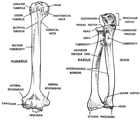
Our primary focus here is the humeroradial joint. This joint, unlike the purely hinge-like humeroulnar joint, allows for a greater range of motion. But which specific part of the radius articulates with the humerus? The answer is the radial head.
The Radial Head: Anatomy and Function
The radial head is the proximal (closest to the body) end of the radius. It's a disc-shaped structure with several key features:
- Articular Surface: This smooth, concave surface is the primary articulating area with the humerus. Its shape perfectly complements the corresponding capitulum of the humerus, allowing for smooth, congruent articulation. This concave shape is crucial for allowing the radius to rotate around the ulna.
- Neck: A constricted area distal to the head, connecting the head to the body of the radius. This neck is a common site for fractures, especially in children.
- Radial Tuberosity: A roughened projection on the medial aspect of the radial neck, serving as an attachment site for the biceps brachii muscle. This muscle plays a significant role in elbow flexion and forearm supination.
The articular surface of the radial head is meticulously designed to fit the slightly convex capitulum of the humerus, facilitating smooth movement during flexion, extension, and pronation/supination. The congruency of these surfaces minimizes friction and maximizes stability.
The Capitulum of the Humerus: The Radius's Partner
To fully understand the humeroradial articulation, we need to examine the humerus's contribution: the capitulum. This is a rounded, lateral projection on the distal end of the humerus. Its smooth, convex surface is perfectly complementary to the concave articular surface of the radial head.
The capitulum's smooth, rounded shape facilitates the gliding movement of the radial head during pronation and supination. Its position relative to the trochlea (the articulation surface for the ulna) ensures coordinated movement of both the radius and ulna during elbow movements.
Mechanics of the Humeroradial Joint
The humeroradial joint's mechanics are intertwined with those of the humeroulnar and proximal radioulnar joints. While the humeroulnar joint primarily governs flexion and extension, the humeroradial joint contributes to these movements and adds the crucial element of rotation:
- Flexion and Extension: The radial head glides along the capitulum during flexion and extension. This movement is facilitated by the concave-convex relationship between the articulating surfaces.
- Pronation and Supination: These movements involve rotation of the radius around the ulna. The radial head pivots on the capitulum, maintaining contact while the radius rotates. This rotation is primarily facilitated by the proximal radioulnar joint, but the humeroradial joint plays a crucial stabilizing role.
Clinical Significance: Injuries and Conditions Affecting the Humeroradial Joint
Understanding the anatomy and function of the humeroradial joint is crucial for diagnosing and managing various clinical conditions. Injuries to this joint are relatively common, particularly:
- Radial Head Fractures: These are frequent injuries, especially among adults, often resulting from a fall onto an outstretched hand. The location and severity of the fracture will determine the treatment strategy.
- Dislocations: The radial head can dislocate from the capitulum, often accompanied by other elbow injuries. Reduction (repositioning) is typically necessary.
- Osteoarthritis: Degeneration of the articular cartilage in the humeroradial joint can lead to pain, stiffness, and limited range of motion.
- Rheumatoid Arthritis: This autoimmune disease can affect the humeroradial joint, causing inflammation, pain, and potential joint destruction.
- Capitellum Fractures: Injuries to the capitulum itself can significantly impact the humeroradial articulation and function of the elbow.
Imaging Techniques for Evaluating the Humeroradial Joint
Various imaging techniques are essential for diagnosing conditions affecting the humeroradial joint. These include:
- X-rays: These provide excellent visualization of bone structures, making them ideal for detecting fractures and dislocations.
- MRI: Magnetic resonance imaging provides detailed images of soft tissues, including ligaments, tendons, and cartilage, allowing for assessment of injuries to these structures and detection of subtle abnormalities.
- CT scans: Computed tomography scans offer detailed cross-sectional images, providing enhanced visualization of bone structures and assisting in the evaluation of complex fractures.
- Ultrasound: This non-invasive imaging method can be used to assess soft tissue structures around the elbow, including muscles and tendons.
These imaging techniques enable accurate diagnosis, guiding appropriate treatment strategies.
Conclusion: A Vital Articulation
The humeroradial joint, with its articulation between the radial head and the capitulum of the humerus, is a vital component of the elbow joint complex. Its unique anatomy and biomechanics allow for a wide range of motion, enabling the precision and dexterity required for many daily tasks. Understanding this specific articulation, its potential injuries, and appropriate diagnostic techniques are fundamental for clinicians and anyone seeking a comprehensive understanding of human anatomy and musculoskeletal function. The intricacy and functionality of the elbow, and specifically the contribution of the humeroradial joint, underscore the remarkable design and engineering of the human body. Further research and advancements in both understanding and treatment of injuries to this region continue to improve outcomes for patients suffering from related pathologies.
Latest Posts
Latest Posts
-
Do Plane Mirrors Form Real Images
Apr 03, 2025
-
After 1880 European Colonization Was Motivated By The
Apr 03, 2025
-
Gaps Or Interruptions In The Myelin Sheath Are Called
Apr 03, 2025
-
Some Bacteria Have Small Extrachromosomal Pieces Of Circular Dna Called
Apr 03, 2025
-
How To Calculate The Bandwidth Of A Signal
Apr 03, 2025
Related Post
Thank you for visiting our website which covers about What Part Of The Radius Articulates With The Humerus . We hope the information provided has been useful to you. Feel free to contact us if you have any questions or need further assistance. See you next time and don't miss to bookmark.
