What Part Of The Eye Has The Greatest Visual Acuity
News Leon
Mar 24, 2025 · 6 min read
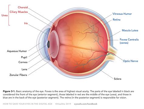
Table of Contents
What Part of the Eye Has the Greatest Visual Acuity?
Visual acuity, the ability to see fine detail, is a crucial aspect of our vision. Understanding which part of the eye contributes most to this sharpness is key to appreciating the complexity and brilliance of our visual system. While the entire visual pathway plays a role, the fovea within the retina is undeniably the champion of visual acuity. This article delves deep into the anatomy and physiology of the fovea, explaining why it reigns supreme in sharp vision, and exploring related factors that influence our overall visual experience.
The Retina: The Eye's Image Sensor
Before focusing on the fovea, let's understand the retina's role. The retina, a light-sensitive tissue lining the back of the eye, is essentially the eye's equivalent of a digital camera sensor. It converts light into electrical signals that are transmitted to the brain via the optic nerve, allowing us to perceive images. The retina is composed of various cell types, including photoreceptor cells (rods and cones), bipolar cells, ganglion cells, and horizontal and amacrine cells. These cells work in concert to process visual information.
Rods and Cones: The Light Receptors
Within the retina, we find two types of photoreceptor cells: rods and cones. Rods are responsible for vision in low-light conditions, providing us with our night vision. They are not involved in high visual acuity. Cones, on the other hand, are crucial for color vision and visual acuity in bright light conditions. They come in three types, each sensitive to different wavelengths of light (red, green, and blue), allowing us to perceive a wide spectrum of colors.
The Fovea: The High-Resolution Center
The fovea, a small, specialized area in the center of the retina, is where visual acuity reaches its peak. Its unique structure and cellular composition are the primary reasons for its superior performance.
Anatomy of the Fovea
The fovea is characterized by several key anatomical features that contribute to its high acuity:
-
High Cone Density: The fovea boasts an extraordinarily high density of cones, significantly more than any other region of the retina. This densely packed arrangement of cones allows for the precise spatial resolution needed for detailed vision. The absence of rods in the fovea's center further enhances cone-mediated visual acuity.
-
Absence of Ganglion Cells: In most parts of the retina, ganglion cells' axons and their supporting cells layer themselves over the photoreceptors. This layer scatters light, reducing visual clarity. However, in the fovea, the ganglion cells and their inner plexiform layer are displaced laterally, minimizing light scattering. This avoids obscuring the light reaching the cones. This unique arrangement ensures that light reaches the cones directly, improving the clarity of the image.
-
Thinning of the Retinal Layers: The overall thinning of the retinal layers in the fovea further reduces light scattering and ensures that light reaches the cones with minimal obstruction. This minimizes blurring and maximizes the sharpness of the image.
-
Specialized Cone Type: The cones in the fovea are predominantly of the M-cone type which are highly sensitive to yellow-green light. This cone type is known for its high spatial resolution contributing to our fine-detail vision.
Why the Fovea is Crucial for Visual Acuity
The combination of these anatomical features in the fovea results in several key advantages for visual acuity:
-
High Spatial Resolution: The high density of cones allows the fovea to resolve fine details. Each cone in the fovea has a dedicated pathway to the brain, leading to a precise one-to-one mapping of light information.
-
Reduced Light Scattering: The displacement of ganglion cells and thinning of retinal layers minimize light scattering, improving image clarity. This ensures sharp, crisp vision, particularly in well-lit conditions.
-
Enhanced Color Vision: While all cones contribute to color vision, the high concentration of cones in the fovea makes it crucial for perceiving fine details in color.
-
Precise Visual Detail: The direct pathway from cones to the brain ensures the faithful transmission of visual information. The brain receives a highly detailed representation of the central visual field.
-
Macular Degeneration and Fovea: The vulnerability of the fovea is highlighted in conditions like Age-related Macular Degeneration (AMD), a leading cause of vision loss in older adults. AMD primarily affects the macula (the area surrounding the fovea) resulting in significant impairment of central vision and decreased visual acuity. This underscores the fovea's critical role in sharp vision.
Beyond the Fovea: The Peripheral Vision's Role
While the fovea reigns supreme in visual acuity, it's important to note the contribution of peripheral vision. Our peripheral vision, handled by the retinal regions outside the fovea, is less sharp but crucial for detecting motion, estimating overall scene layout, and maintaining spatial awareness. The lower density of cones and presence of rods in the peripheral retina allow for broader field of view and increased light sensitivity, which are crucial for overall visual functionality and safety.
Factors Affecting Visual Acuity
While the fovea is the primary determinant of visual acuity, several other factors play a significant role:
-
Refractive Errors: Conditions like myopia (nearsightedness), hyperopia (farsightedness), and astigmatism can blur the image focused on the retina, reducing overall visual acuity. Corrective lenses are often necessary to address these issues.
-
Neural Processing: The brain's interpretation of retinal signals also influences our perception of visual acuity. Neurological conditions can impact how the brain processes visual information, impacting visual acuity despite a healthy retina.
-
Optical Quality: The clarity of the eye's optical components—the cornea and lens—significantly influences the quality of the image projected onto the retina. Cataracts, for example, can dramatically reduce visual acuity.
-
Lighting Conditions: Adequate lighting is essential for optimal cone function and, therefore, sharp vision. Dim lighting heavily favors the rods over the cones, severely impacting visual acuity.
Conclusion: The Fovea's Undisputed Reign
In conclusion, while the entire visual system contributes to our perception of the world, the fovea within the retina stands out as the key player in achieving high visual acuity. Its unique anatomical structure—high cone density, absence of overlying ganglion cells, and thinning of retinal layers—allows for superior spatial resolution, minimized light scattering, and precise transmission of visual information to the brain. Understanding the fovea's vital role underscores the complexity and wonder of our visual system and highlights its importance in our daily lives. Maintaining eye health and addressing any refractive errors is crucial to preserve the remarkable ability of the fovea to provide us with sharp, detailed vision. The intricate interplay between the fovea and the broader visual pathway reminds us of the remarkable engineering of the human eye, a masterpiece of evolution that allows us to experience the richness and detail of the visual world.
Latest Posts
Related Post
Thank you for visiting our website which covers about What Part Of The Eye Has The Greatest Visual Acuity . We hope the information provided has been useful to you. Feel free to contact us if you have any questions or need further assistance. See you next time and don't miss to bookmark.