What Is The Measure Of Acd
News Leon
Apr 04, 2025 · 6 min read

Table of Contents
What is the Measure of ACD? Understanding Angle-Closure Glaucoma and its Diagnosis
Angle-closure glaucoma (ACD), also known as narrow-angle glaucoma, is a serious condition affecting the eye's drainage system. Understanding the "measure" of ACD isn't about a single numerical value, but rather a comprehensive assessment of several factors contributing to the risk and severity of this condition. This article delves into the intricacies of measuring and managing ACD, encompassing diagnostic techniques, risk factors, and treatment options.
Understanding the Anatomy of Angle Closure
Before we delve into the measurement aspects, let's clarify the anatomy involved. The eye's drainage system, responsible for removing aqueous humor (the fluid that nourishes the eye), is crucial. This system involves the trabecular meshwork and Schlemm's canal. In angle-closure glaucoma, the angle between the iris (the colored part of the eye) and the cornea (the clear front part of the eye) is narrowed, obstructing the drainage pathway. This build-up of pressure, known as intraocular pressure (IOP), can damage the optic nerve, leading to vision loss.
The Anterior Chamber Angle: The Key Player
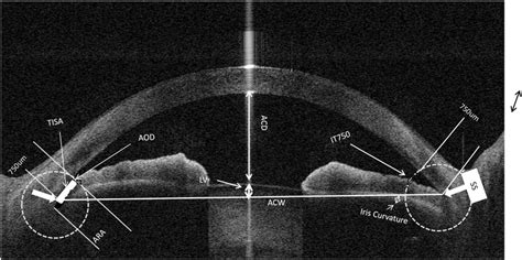
The anterior chamber angle (ACA) is the crucial area where the iris meets the cornea. Its width and openness directly impact the eye's drainage capacity. A narrow angle increases the risk of angle closure. Measuring this angle is central to diagnosing and managing ACD.
Measuring the Anterior Chamber Angle: Diagnostic Techniques
There are several methods ophthalmologists use to assess the ACA and determine the severity of angle closure. These methods vary in their invasiveness and the information they provide.
1. Gonioscopy: The Gold Standard
Gonioscopy is considered the gold standard for ACA assessment. It's a non-invasive procedure where a special lens is placed on the eye's surface, allowing the ophthalmologist to visualize the angle. The gonioscopist classifies the angle based on the structures visible and their relationship to each other. This provides a detailed picture of the angle's anatomy and helps determine the degree of closure. Different grading systems exist, but the fundamental goal is to assess the openness of the angle and identify any potential blockages.
Types of Gonioscopic Findings:
- Open angle: The angle is wide open, with ample space for aqueous humor drainage. This indicates a low risk of angle-closure glaucoma.
- Narrow angle: The angle is partially closed, indicating a higher risk of angle closure.
- Closed angle: The angle is completely closed, preventing aqueous humor drainage and causing a rapid increase in IOP. This is a critical situation requiring immediate intervention.
2. Ultrasonography (UBM): A Detailed Look
Ultrasound biomicroscopy (UBM) provides a more detailed image of the ACA than gonioscopy. This imaging technique utilizes high-frequency sound waves to create detailed cross-sectional images of the eye's anterior segment, revealing the angle's anatomy with remarkable precision. UBM is particularly useful in evaluating the structures within the angle, such as the iris, ciliary body, and trabecular meshwork, which can contribute to angle closure.
3. Pachymetry: Assessing Corneal Thickness
While not a direct measure of the ACA, corneal thickness plays a crucial role in IOP measurement. Pachymetry measures the thickness of the cornea. A thicker cornea can lead to inaccurate IOP readings, potentially masking or overestimating the severity of ACD.
4. Intraocular Pressure (IOP) Measurement: A Crucial Indicator
Measuring IOP is paramount in diagnosing and managing glaucoma, including ACD. Elevated IOP is a key indicator of the condition. Various methods are used, including tonometry, which involves using an instrument to gently flatten the cornea and measure the resistance. This provides a numerical value for IOP, usually expressed in millimeters of mercury (mmHg). While IOP is a critical factor, it's not the sole determinant of glaucoma; other factors, such as the ACA, optic nerve damage, and visual field loss, must be considered.
Risk Factors for Angle-Closure Glaucoma
Several factors increase the risk of developing angle-closure glaucoma. Understanding these risk factors is essential for early detection and prevention.
1. Age and Ethnicity: A Predisposing Factor
Angle-closure glaucoma is more common in older individuals and certain ethnic groups, particularly those of East Asian descent. As people age, the lens within the eye tends to thicken, potentially further narrowing the ACA.
2. Hyperopia (Farsightedness): A Structural Influence
Hyperopic eyes (farsighted) tend to have a shallower anterior chamber, making them more susceptible to angle closure. The shorter axial length increases the likelihood of iris crowding, diminishing the drainage pathway.
3. Family History: Genetic Predisposition
A family history of angle-closure glaucoma significantly increases the risk. This suggests a genetic component influencing the eye's anatomy and predisposition to this condition.
4. Certain Medications: Potential Side Effects
Some medications, particularly those affecting the autonomic nervous system, can influence pupil size and potentially contribute to angle closure.
5. Eye Trauma: A Physical Impact
Eye injuries can disrupt the eye's normal anatomy, potentially leading to angle closure.
Managing and Treating Angle-Closure Glaucoma
Treatment for angle-closure glaucoma depends on the severity of the condition and the presence of acute symptoms.
1. Laser Peripheral Iridotomy (LPI): A Minimally Invasive Procedure
LPI is a common procedure for managing ACD. A small laser hole is created in the iris, allowing aqueous humor to flow freely and relieving pressure build-up. This procedure effectively prevents acute angle closure episodes.
2. Medications: Lowering Intraocular Pressure
Medications, such as eye drops that lower IOP, are often prescribed to manage the condition. These medications can help to reduce the pressure within the eye, protecting the optic nerve.
3. Surgery: Addressing Severe Cases
In severe cases where medication and LPI are insufficient, surgery may be necessary. Various surgical techniques can address angle closure and improve aqueous humor drainage.
The Importance of Regular Eye Exams
Regular comprehensive eye exams are crucial for early detection of ACD. Early diagnosis and intervention can significantly reduce the risk of vision loss. During these exams, the ophthalmologist will assess IOP, perform gonioscopy or other imaging techniques, and evaluate the optic nerve and visual fields.
Conclusion: A Holistic Approach to Measuring ACD
"Measuring" ACD isn't a single metric; it's a comprehensive evaluation combining anatomical assessment (gonioscopy, UBM), physiological measurements (IOP), risk factor analysis, and consideration of the patient's overall health. Early detection through regular eye examinations and proactive management strategies—including laser procedures or medications—are vital for preserving vision and improving the quality of life for individuals affected by angle-closure glaucoma. The collaborative efforts of the patient and their ophthalmologist are crucial for effective management and successful outcomes. Remember, proactive care significantly improves the chances of preserving eyesight and preventing the devastating effects of ACD.
Latest Posts
Latest Posts
-
The Light Reactions Of Photosynthesis Occur In The
Apr 04, 2025
-
A Body At Rest Can Have
Apr 04, 2025
-
Router Operate At Which Layer Of The Osi Model
Apr 04, 2025
-
Which Of The Following Colors Has The Longest Wavelength
Apr 04, 2025
-
The Cell Membrane Is Composed Mainly Of
Apr 04, 2025
Related Post
Thank you for visiting our website which covers about What Is The Measure Of Acd . We hope the information provided has been useful to you. Feel free to contact us if you have any questions or need further assistance. See you next time and don't miss to bookmark.
