The Ends Of A Long Bone Are Called The
News Leon
Mar 14, 2025 · 7 min read
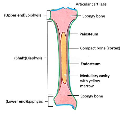
Table of Contents
The Ends of a Long Bone are Called the Epiphyses: A Deep Dive into Bone Anatomy and Development
The ends of a long bone are called the epiphyses (singular: epiphysis). Understanding the epiphyses is crucial to comprehending bone growth, development, and various skeletal conditions. This comprehensive article will delve into the intricate structure and function of the epiphyses, exploring their role in skeletal maturation and highlighting their clinical significance.
What are Epiphyses?
Epiphyses are the secondary ossification centers located at the ends of long bones. Unlike the diaphysis (the shaft of the long bone), which ossifies first, the epiphyses begin their ossification process later in development. This secondary ossification is vital for longitudinal bone growth. The epiphyses are primarily composed of spongy bone, also known as cancellous bone, a porous and less dense type of bone tissue compared to the compact bone found in the diaphysis. This spongy structure provides strength while maintaining a relatively low weight. The epiphyses are covered by a layer of articular cartilage, which allows for smooth, low-friction movement at the joints.
Key Characteristics of Epiphyses:
- Spongy Bone Structure: The internal structure of the epiphyses is characterized by a network of trabeculae, which are thin, bony plates arranged in a lattice-like pattern. This architecture maximizes strength and minimizes weight.
- Red Bone Marrow: The spaces within the trabeculae of the epiphyses contain red bone marrow, which is responsible for the production of blood cells (hematopoiesis).
- Articular Cartilage: A thin layer of hyaline cartilage covers the articular surfaces of the epiphyses, providing a smooth, lubricated surface that minimizes friction during joint movement. This cartilage is essential for joint health and prevents wear and tear.
- Epiphyseal Plate (Growth Plate): In growing bones, a crucial structure called the epiphyseal plate, or growth plate, separates the epiphysis from the diaphysis. This plate is composed of actively dividing cartilage cells that contribute to the lengthening of the bone.
The Role of Epiphyses in Bone Growth: The Epiphyseal Plate
The epiphyseal plate, also known as the growth plate, is a cartilaginous structure located between the epiphysis and the metaphysis (the widening portion of the diaphysis near the epiphysis). It's the primary site of longitudinal bone growth. The epiphyseal plate consists of several zones of actively proliferating and differentiating chondrocytes (cartilage cells):
Zones of the Epiphyseal Plate:
- Zone of Resting Cartilage: This zone anchors the epiphyseal plate to the epiphysis. The chondrocytes here are relatively inactive.
- Zone of Proliferation: Chondrocytes in this zone undergo rapid cell division, increasing the length of the epiphyseal plate.
- Zone of Hypertrophy: The chondrocytes mature and enlarge in this zone, accumulating glycogen and lipids.
- Zone of Calcification: The extracellular matrix surrounding the hypertrophic chondrocytes calcifies, preparing the cartilage for ossification.
- Zone of Ossification: Osteoblasts invade the calcified cartilage, depositing bone matrix and replacing the cartilage with bone tissue.
This continuous process of cartilage proliferation, maturation, calcification, and ossification leads to an increase in the length of the bone. The rate of growth in the epiphyseal plate is regulated by various factors, including hormones like growth hormone, thyroid hormone, and sex hormones.
Closure of the Epiphyseal Plate: The Epiphyseal Line
As individuals reach skeletal maturity, the rate of cartilage production in the epiphyseal plate slows down, eventually ceasing completely. This process is known as epiphyseal plate closure. The epiphyseal plate is then replaced by bone tissue, forming a bony structure called the epiphyseal line. The closure of the epiphyseal plates marks the end of longitudinal bone growth. The timing of epiphyseal plate closure varies depending on the bone and the individual's genetics and overall health.
Clinical Significance of Epiphyses and Epiphyseal Plates:
Understanding the epiphyses and their development is critical in various clinical settings:
Fractures:
Epiphyseal fractures are common injuries, particularly in children and adolescents whose epiphyseal plates are still open. These fractures can disrupt bone growth and may lead to deformities if not treated properly. The Salter-Harris classification system is used to categorize epiphyseal fractures based on their location and severity.
Epiphyseal Dysplasia:
This is a group of genetic disorders that affect the development of the epiphyses, leading to abnormalities in bone growth and skeletal proportions. These disorders can cause dwarfism or other skeletal deformities.
Osteoarthritis:
The articular cartilage covering the epiphyses plays a crucial role in joint health. Degeneration of this cartilage, as seen in osteoarthritis, leads to pain, stiffness, and reduced joint mobility.
Bone Tumors:
Epiphyses can be affected by both benign and malignant bone tumors. The location of the tumor within the bone influences the diagnosis and treatment strategy.
Bone Infections (Osteomyelitis):
Infections can spread to the epiphyses through the bloodstream or directly from a nearby joint. Early diagnosis and treatment are essential to prevent complications.
The Importance of Understanding Epiphyses:
The epiphyses are not merely the ends of long bones; they are dynamic structures crucial for skeletal development, growth, and overall health. Their role in longitudinal bone growth, blood cell production, and joint function underscores their importance. Clinically, understanding epiphyseal development and potential pathologies is vital for the diagnosis and management of a wide range of skeletal conditions. From pediatric fractures to adult-onset osteoarthritis, the epiphyses are central to understanding the complexities of the skeletal system.
Beyond Long Bones: Epiphyses in Other Bone Types
While the term "epiphysis" is most often associated with long bones, the concept of secondary ossification centers extends to other bone types. While not perfectly analogous to the epiphyses in long bones, these secondary ossification centers contribute to the overall development and shape of the bone. For example, short bones like those in the wrist and ankle also have secondary ossification centers that contribute to their final shape. Flat bones, like those in the skull, also develop through intramembranous ossification, which involves the formation of bone directly from mesenchymal tissue, without a cartilage intermediate. While these processes differ slightly from the endochondral ossification seen in long bones, the underlying principle of secondary ossification centers remains relevant.
The Interplay of Genetics, Hormones, and Nutrition in Epiphyseal Development
The development and maturation of the epiphyses are influenced by a complex interplay of genetic, hormonal, and nutritional factors. Genetic factors determine the overall skeletal plan and the timing of various developmental milestones. Hormones, including growth hormone, thyroid hormone, and sex hormones, play crucial roles in regulating the rate of cartilage proliferation and ossification in the epiphyseal plate. Nutritional factors, such as adequate calcium and vitamin D intake, are essential for proper bone mineralization and overall skeletal health. Any disruption in these factors can significantly impact epiphyseal development and potentially lead to various skeletal abnormalities.
Imaging Techniques for Assessing Epiphyseal Development and Pathology
Various imaging techniques are utilized to assess epiphyseal development and identify potential pathologies. X-rays are commonly used to visualize bone structure and assess the status of the epiphyseal plates. Magnetic resonance imaging (MRI) provides detailed images of soft tissues, making it useful for evaluating the articular cartilage and other surrounding structures. Computed tomography (CT) scans offer high-resolution images of bone, which can be helpful in identifying subtle fractures or other bone abnormalities. These imaging techniques are essential tools for diagnosing and monitoring various skeletal conditions involving the epiphyses.
Conclusion: The Enduring Importance of Epiphyseal Study
The epiphyses represent a fascinating and crucial aspect of bone biology. Their role in bone growth, joint function, and overall skeletal health is undeniable. Furthermore, a thorough understanding of epiphyseal development and potential pathologies is paramount for clinicians involved in the diagnosis and management of a wide range of skeletal conditions. From childhood injuries to adult-onset diseases, the epiphyses remain a central focus in the ongoing study of bone biology and skeletal health. Continued research in this area promises to further elucidate the complexities of epiphyseal development and provide improved diagnostic and therapeutic strategies for skeletal conditions affecting the epiphyses and the broader skeletal system.
Latest Posts
Latest Posts
-
Which Of The Following Needs A Proof
Mar 14, 2025
-
Pure Substances Are Made Of Only One Type Of
Mar 14, 2025
-
A Dpt Vaccination Is An Example Of
Mar 14, 2025
-
Whats The Difference Between An Enzyme And A Hormone
Mar 14, 2025
-
An Alloy With One Of The Constituents Being Mercury
Mar 14, 2025
Related Post
Thank you for visiting our website which covers about The Ends Of A Long Bone Are Called The . We hope the information provided has been useful to you. Feel free to contact us if you have any questions or need further assistance. See you next time and don't miss to bookmark.
